A kit and detection method for detecting human regulatory T cell subtypes
A technology for human detection and regulation, applied in biological testing, preparation of test samples, measuring devices, etc., can solve problems such as inability to further identify CD4
- Summary
- Abstract
- Description
- Claims
- Application Information
AI Technical Summary
Problems solved by technology
Method used
Image
Examples
Embodiment 1
[0090] To isolate mononuclear cells from whole blood:
[0091] 1) Take 1mL of fresh venous blood (blood isolated for ≤2h) and add 1mL of blood thinner to dilute the blood;
[0092] 2) Take 1mL of mononuclear cell separation solution at the bottom of the centrifuge tube, and carefully superimpose the diluted blood on top of the mononuclear cell separation solution to avoid mixing of blood and mononuclear cell separation solution;
[0093] 3) In the centrifuge, centrifuge at 800g for 20 minutes, set the temperature of the centrifuge to 18°C, set the speed up to 9, and set the speed down to 0;
[0094] 4) Use a dropper to suck out the buffy coat (mononuclear cells) on the interface, and try not to suck out the upper layer of liquid;
[0095] 5) Dilute the mononuclear cell solution with 2 mL of blood diluent, centrifuge at 250 g at room temperature for 10 min; collect the cell pellet, resuspend the mononuclear cells in 100 μL of cell culture medium, and obtain the mononuclear cel...
Embodiment 2
[0097] Stimulation of human peripheral blood mononuclear cells:
[0098] 1) Count the cells, adjust the cell density to 1×10 with cell culture medium 6 / mL;
[0099] 2) For every 1 mL of cell suspension, add 2 μL of lymphocyte activation solution and 1 μL of LPS, mix evenly, inoculate on a cell culture plate, and place in CO 2 Incubate at 37°C for 4 hours in an incubator;
[0100] 3) Collect 1 mL of cells in each tube, add 1 mL of phosphate buffer, and centrifuge at 200 g for 6 min;
[0101] 4) Count the cells, adjust the cell density to 1×10 with phosphate buffer 7 / mL cell suspension.
Embodiment 3
[0103] Fluorescent antibody staining:
[0104] 1) Add 100μL cell suspension to each tube, then add 1uL dissolved dead cell removal dye and 5uL FcR blocker, and incubate at room temperature for 15min in the dark;
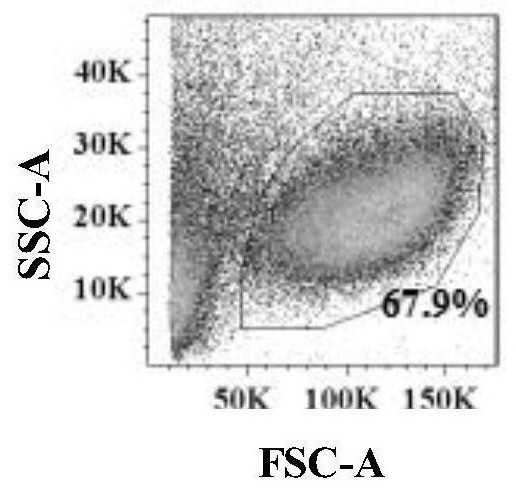
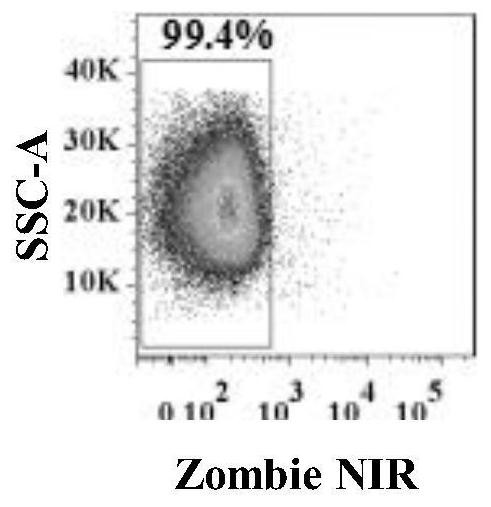
[0105] 2) Add 1mL cell staining buffer to each tube, centrifuge at 200g for 6min, and discard the supernatant; the obtained human peripheral blood mononuclear cell population after removing dead cells is as follows: figure 2 shown;
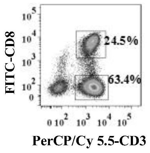
[0106] 3) Add 100 μL cell staining buffer to each tube to resuspend the cells, then add PerCP / Cy 5.5-labeled anti-human CD3, FITC-labeled anti-human CD8, PE-labeled anti-human CD25, BrilliantViolet 421-labeled anti-human TGF-β and BrilliantViolet 510-labeled anti-human CD127 antibody 5uL each, or PerCP / Cy 5.5-labeled anti-human CD3, FITC-labeled anti-human CD8, BrilliantViolet 510-labeled anti-human CD25, PE / Cy7-labeled anti-human CD278 (ICOS) and PE Incubate 5uL of each labeled anti-human CCR6 antibody at room temperature in the dark...
PUM
| Property | Measurement | Unit |
|---|---|---|
| osmolarity | aaaaa | aaaaa |
Abstract
Description
Claims
Application Information
 - R&D
- Intellectual Property
- Life Sciences
- Materials
- Tech Scout
- Unparalleled Data Quality
- Higher Quality Content
- 60% Fewer Hallucinations
Browse by: Latest US Patents, China's latest patents, Technical Efficacy Thesaurus, Application Domain, Technology Topic, Popular Technical Reports.
© 2025 PatSnap. All rights reserved.Legal|Privacy policy|Modern Slavery Act Transparency Statement|Sitemap|About US| Contact US: help@patsnap.com


