Micro-fluidic device and method for negative separation of circulating tumor cells and clusters

A microfluidic device and tumor cell technology, applied in the field of microfluidic devices for negative separation of circulating tumor cells and clusters, can solve the problems of scarcity of CTCs and CTC-Clusters, difficulty in simultaneous separation, and heterogeneity, etc. Achieve the effect of high sensitivity, low cost and short time consumption

Inactive Publication Date: 2018-10-19
UNIV OF SCI & TECH BEIJING
View PDF7 Cites 7 Cited by
  • Summary
  • Abstract
  • Description
  • Claims
  • Application Information

AI Technical Summary

Problems solved by technology

At present, many CTCs separation methods can only achieve the separation of CTCs or CTC-Clusters using microfluidic technology, and it is difficult to efficiently separate the two simultaneously.
At present, the identification of CTCs mainly relies on epithelial markers such as EpCAM and CK, and it is difficult to identify CTCs from EMT transition process and non-epithelial sources
Moreover, CTCs and CTC-Clusters in blood are rare and heterogeneous, so it is urgent to develop an efficient and simultaneous separation method for CTCs and CTC-Clusters and a detection method that can better identify all CTCs

Method used


Image

  • Micro-fluidic device and method for negative separation of circulating tumor cells and clusters
  • Micro-fluidic device and method for negative separation of circulating tumor cells and clusters
  • Micro-fluidic device and method for negative separation of circulating tumor cells and clusters

Examples


Embodiment 1

[0046] Example 1: Separation, screening and precise positioning of single circulating tumor cells using a microfluidic device

[0047] (1) Pre-treatment of the separation device: For the separation device, its structure is as follows figure 1 shown. A mixed solution of 10% goat serum and 3% bovine serum albumin was injected into the solution storage chamber and closed for 1 hour to avoid non-specificity of cells.

[0048] (2) Negative enrichment of tumor cells: First, take 4ml of cancer patient blood sample and dilute it, then centrifuge at low speed to discard the upper layer of serum and platelets, and set aside for use; secondly, after density gradient centrifugation of the sample processed in the previous step, absorb the middle nucleated cell layer; Again, after co-incubating the samples treated in the previous step with CD45 and CD15 antibody-labeled immunomagnetic beads, white blood cells were removed by a magnetic frame to obtain tumor cell-enriched samples. Finally,...

Embodiment 2

[0052] Example 2: Rapid Isolation of Circulating Tumor Cell Clusters in Peripheral Blood

[0053] (1) Pretreatment of the separation device: For the separation device, a mixed solution of 10% goat serum and 3% bovine serum albumin was injected into the solution storage chamber to seal for 1 hour to avoid non-specificity of cells.

[0054] (2) Preparation of simulated cancer patient blood samples containing tumor cell clusters: 4 ml of peripheral blood samples from healthy volunteers were collected with an anticoagulant tube, and several tumor cell clusters of LOVO cell lines were selected with capillary tubes and mixed into blood to simulate cancer patient blood samples.

[0055] (3) Negative enrichment of tumor cell clusters: First, after dilution of the sample, centrifuge at low speed to discard the upper layer of serum and platelets for use; secondly, after density gradient centrifugation of the sample processed in the previous step, absorb the nucleated cell layer in the mi...

Embodiment 3

[0059] Example 3: Individual isolation and purification of tumor cell clusters after screening and precise positioning

[0060] (1) Rapid separation of tumor cell clusters in peripheral blood (the steps are the same as in Example 2).

[0061] (2) Separation and purification: Under bright field, find the micropits after screening and precise positioning, and use capillary to purify and separate circulating tumor cell clusters. After picking, reconfirmation was performed in brightfield and fluorescent field.

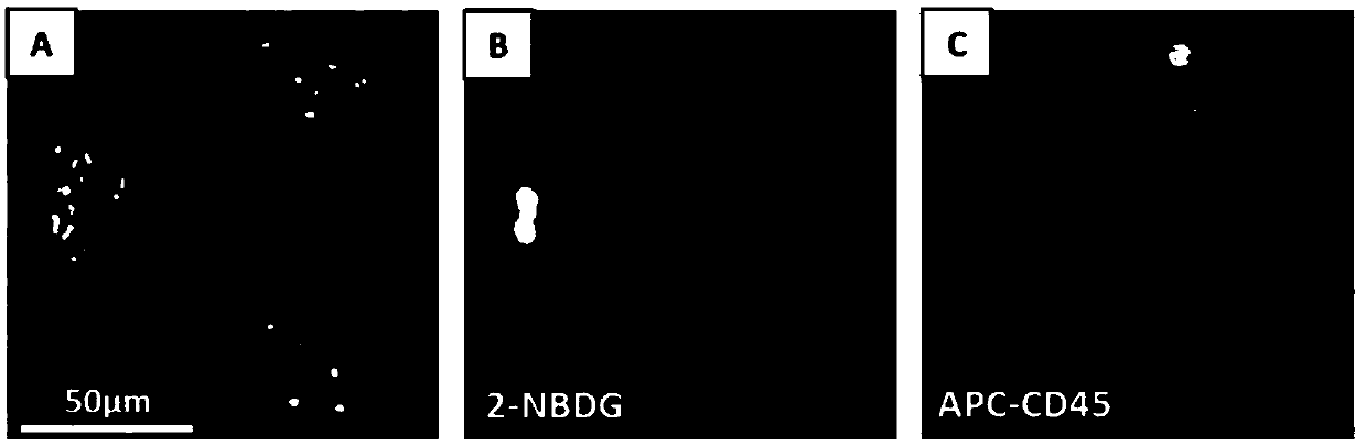
[0062] Figure 4 Middle A is a bright-field microscope view of the 21E-labeled array under a 20X magnification, which can precisely locate tumor cell clusters in (21, E, 6 rows, 6 rows) micropits. in Figure 4 Middle B is the field of view of the 21E-marked array under a 20X magnification microscope after the capillary needle is selected. It can be seen (21, E, 6 rows, 6 rows) that the tumor cell clusters in the micropits have been separated. in Figure 4 Middle C is a ...


PUM


Abstract

The invention provides a micro-fluidic device for negative separation of circulating tumor cells and clusters. The micro-fluidic device is made by assembling a sample storage chamber and a micro-fluidic chip by virtue of thermal bonding, the micro-fluidic device has the diameter of 60mm and is the same as a cell-culture dish having the diameter of 60mm, and the micro-fluidic device can be just putinto the culture dish to centrifuge. A micro-cavity array chip is capable of synchronously capturing CTCs (Circulating Tumor Cells) and CTCs-Clusters, and on the base of the micro-fluidic device, synchronous negative separation of the CTCs and CTCs-Clusters can be realized; the whole micro-cavity array chip is divided into 64 regions by numbers from 1 to 64, and each region is divided into 16 cells by letters from A to P, so that each micro cavity can be accurately located, accurate localization and single-cell isolation purification of the enriched CTCs / CTCs-Clusters can be realized, and convenience is provided for performing single-cell analysis on subsequent target cells.

Description

technical field [0001] The invention belongs to the technical field of biological and medical detection, and in particular relates to a microfluidic device and method for negative separation of circulating tumor cells and clusters. Background technique [0002] Circulating tumor cells (CTCs) are shed from the primary tumor site, enter the blood circulation after epithelial-mesenchymal transition, and can be located in remote sites, proliferate and form new lesions after mesenchymal-epithelial transition occurs. Circulating tumor cell clusters (CTC-clusters) refer to cell clusters formed by the aggregation of multiple CTCs of the same kind or with other types of cells. Circulating tumor cells and circulating tumor cell clusters in the blood are closely related to cancer metastasis. Simultaneous detection of CTCs and CTC-Clusters is beneficial to early diagnosis of cancer, evaluation of curative effect, and research on the mechanism of tumor metastasis. [0003] Clinical stud...

Claims


Application Information

Patent Timeline
Patent Type & Authority Applications(China)
IPC IPC(8): B01L3/00
CPCB01L3/5027B01L3/50273B01L2300/0861B01L2300/12B01L2400/0409
Inventor 赵亮蒋海祥刘杨张学记
Owner UNIV OF SCI & TECH BEIJING
Why Eureka
Try Eureka