A gold nanopore array-based surface-enhanced Raman scattering detection method for DNA methylation and its application
A surface-enhanced Raman and detection method technology, which is applied in Raman scattering, measuring devices, and material analysis through optical means, can solve problems such as complex amplification, labeling procedures, large amount of DNA, and unstable measurement. Simple operation, high reproducibility, good Raman enhancement effect
- Summary
- Abstract
- Description
- Claims
- Application Information
AI Technical Summary
Problems solved by technology
Method used
Image
Examples
Embodiment 1
[0044] Preparation of basic solution
[0045] 1. Preparation of gold nanopore arrays
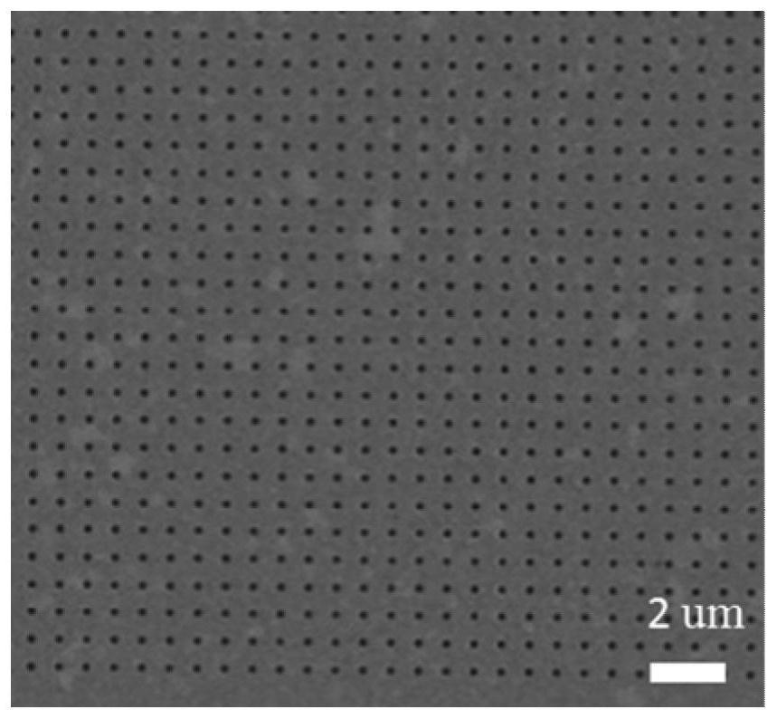
[0046] Gold nanohole arrays were prepared using an electron beam exposure system and thermal evaporation coating. A 200-nm thick PMMA electron-beam etchant layer was spin-coated on a Si wafer, and then the PMMA-coated substrate was produced under an electron beam with 200-nm apertures and 385-nm edges. 50 μm × 50 μm nanowell array with a distance between them. After developing for 70 seconds in a PMMA developer with a mass ratio of 1:3 methyl isobutyl ketone / isopropanol (MIBK / IPA), a porous structure was produced, followed by IPA rinsing and post-baking at 95°C for 30 minutes, and then in A 50nm-thick gold film was evaporated on the substrate to complete the preparation of the nanopore array. Before use, the substrate was cleaned in UV ozone for 20 minutes with a 18.2MΩ·cm -1 Rinse with deionized water and rinse with N 2 dry. A clean gold nanopore array substrate is obtained. Characteri...
Embodiment 2
[0050] Raman Performance Evaluation of Gold Nanohole Arrays
[0051] The enhancement effect of the gold nanohole array was studied by Raman spectroscopy; a laser of 785nm was selected.
[0052] With the Rh6G solution prepared in Example 1, detect the surface-enhanced Raman spectrum of the solution;
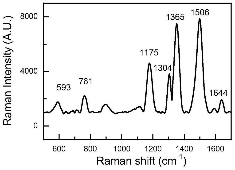
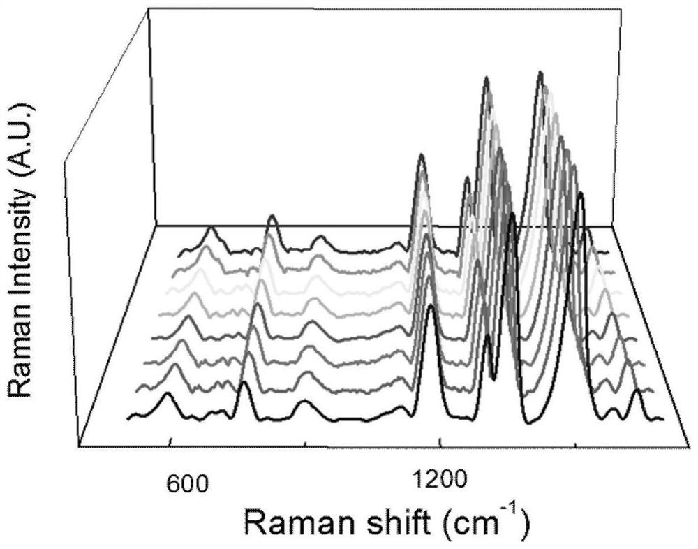
[0053] A piece of gold nanopore array prepared in Example 1 is immersed in the prepared Rh6G solution, and the Raman spectrum of the Rh6G solution is detected by a Raman spectrometer, as shown in figure 2 As shown, the direct detection of 10 at the nanopore -6 Raman signal of mol / L rhodamine 6G. It can be found that there is an obvious Raman enhancement effect in the nanohole area, and the Raman enhancement factor is close to 10 6 . Randomly select different regions in the nanopore array for detection, such as image 3 As shown, it can be found that any region of the nanohole array has a very uniform and stable Raman response, and a Raman signal with good reproducibility can...
Embodiment 3
[0055] Gold Nanopore Arrays for Detecting DNA Methylation
[0056] Take the DNA solution prepared in Example 1 with a concentration of 100 μmol / L and drop-coat it on the gold nanopore array, take it out after 0.5 hours, dry it with nitrogen, and use a Raman instrument with a 785nm laser to detect and study the properties of S1 and S2. Raman spectroscopy. Compare the characteristic differences of their Raman spectra, such as Figure 4 Shown, the characteristic peak of methyl (at 1365cm -1 ) intensity is significantly enhanced, while the characteristic peak of the base cytosine (located at 785cm -1 ) intensity is relatively weakened, and DNA methylation detection can be realized. Take S2 to prepare a set of solutions with different concentrations of DNA, the concentration range is 0.1 μmol / L to 100 μmol / L; the Raman signal of this series of DNA solutions with different concentrations is detected by Raman spectrometer, as shown in Figure 5 As shown, the DNA concentration o...
PUM
| Property | Measurement | Unit |
|---|---|---|
| thickness | aaaaa | aaaaa |
| radius | aaaaa | aaaaa |
Abstract
Description
Claims
Application Information
 - R&D
- Intellectual Property
- Life Sciences
- Materials
- Tech Scout
- Unparalleled Data Quality
- Higher Quality Content
- 60% Fewer Hallucinations
Browse by: Latest US Patents, China's latest patents, Technical Efficacy Thesaurus, Application Domain, Technology Topic, Popular Technical Reports.
© 2025 PatSnap. All rights reserved.Legal|Privacy policy|Modern Slavery Act Transparency Statement|Sitemap|About US| Contact US: help@patsnap.com


