Retina stratification method in eye ground OCT (Optical Coherence Tomography) image

An image center and retinal technology, applied in the field of medical image processing, to achieve the effects of verification accuracy and reliability, strong comparability, good repeatability and adaptability

Active Publication Date: 2018-11-20
杭州富扬科技有限公司
View PDF7 Cites 8 Cited by
  • Summary
  • Abstract
  • Description
  • Claims
  • Application Information

AI Technical Summary

Problems solved by technology

However, graph theory algorithms are very sensitive to noise and image degradation

Method used


Image

  • Retina stratification method in eye ground OCT (Optical Coherence Tomography) image
  • Retina stratification method in eye ground OCT (Optical Coherence Tomography) image
  • Retina stratification method in eye ground OCT (Optical Coherence Tomography) image

Examples


Embodiment Construction

[0054] The present invention will be described in further detail below in conjunction with the accompanying drawings and embodiments. It should be understood that the specific embodiments described here are only used to explain the present invention, not to limit the present invention.

[0055] Embodiments of the present invention are as follows:

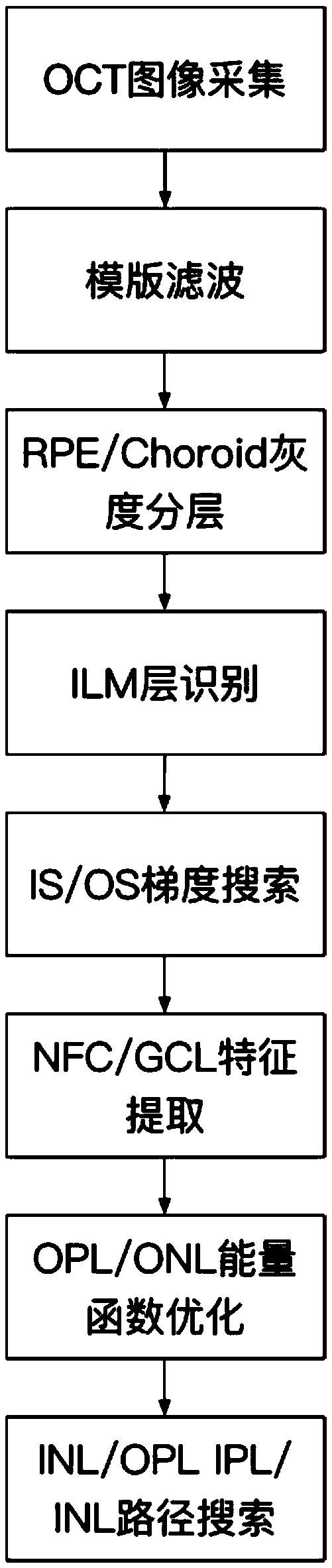
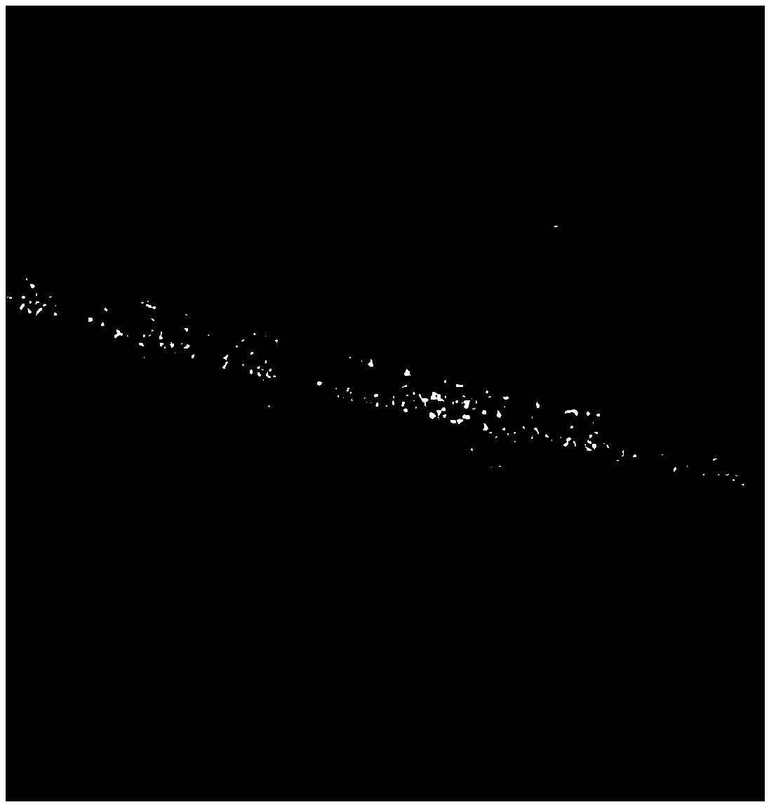
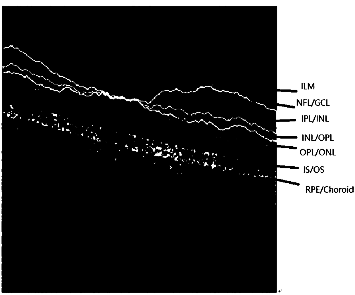
[0056] 1) Collect the OCT image of the fundus retina as the original image, such as figure 2 shown.

[0057] 2) Use the weight coefficient matrix template [1 / 9, 1 / 9, 1 / 9; 1 / 6, 1 / 6, 1 / 6; 1 / 9, 1 / 9, 1 / 9;] to traverse the entire original image for Template filtering;

[0058] 3) Process in the following manner to obtain each boundary line, and perform retinal delamination in the fundus OCT image.

[0059] 3.1) For each column of the OCT image, record the pixel point where the maximum gray value is located, and connect the pixel points where the maximum gray value of each column is located as the dividing line between the retinal pi...


PUM


Abstract

The invention discloses a retina stratification method in an eye ground OCT (Optical Coherence Tomography) image. The eye ground OCT image is acquired as an original image; the integral original imageis traversed by using a weight coefficient matrix template to carry out template filtering; and then RPE (Retina Pigment Epithelium) / Choroid gray scale stratification, ILM (Internal Limiting Membrane) layer identification, IS / OS (Inner Segment / Outer Segment) gradient search, NFC / GCL (Nerve Fiber Layer / Ganglion Cell Layer) feature extraction, OPL / ONL (Outer Molecular Layer / Outer Nuclear Layer) energy function optimization and INL (Inner Nuclear Layer) / OPL and IPL (Inner Molecular Layer) / INL path search are carried out, and finally, segmentation on different layers of the retina in the OCT image is completed. The retina stratification method in the eye ground OCT image, which is disclosed by the invention, can implement automatic segmentation on multiple layer structures by utilizing a computer which is commonly configured, implements effective automatic detection on a complex disease stratified structure of the retina, adopts serialized stratification, has few original processing stepsfor the image, and has a certain advantage in detection efficiency.

Description

technical field [0001] The invention belongs to the field of medical image processing, and relates to a processing method for OCT images, in particular to a retinal layering method in fundus OCT images. Background technique [0002] Optical coherence tomography (OCT) is a high-resolution cross-sectional imaging technique that collects photons of back-reflected light from microstructures on the sample surface and interferes with light from the reference light path to achieve imaging. OCT equipment was first used in the in vivo analysis of biological tissues, mainly in the field of ophthalmology; with the deepening of research, OCT and its images produced by different manufacturers have brought great reference to the clinical diagnosis of ophthalmic diseases. The total thickness of the retinal layer and the thickness of the inner retinal layer are important indicators for clinical ophthalmologists. For example, the thickness of retinal nerve fiber layer (RNFL) and retinal pigm...

Claims


Application Information

Patent Timeline
Patent Type & Authority Applications(China)
IPC IPC(8): A61B3/10A61B3/12G06T7/00G06T7/13
CPCA61B3/102A61B3/12G06T7/0012G06T7/13G06T2207/10101G06T2207/30041
Inventor 周扬陆扬陈正伟刘铁兵陈才施秧
Owner 杭州富扬科技有限公司
Why Eureka
Try Eureka