Automatic retinal vessel segmentation method for glaucoma

A technology for automatic segmentation of retinal blood vessels, applied in image analysis, image enhancement, instruments, etc., can solve the problems of limiting application and promotion, affecting the efficiency of retinal blood vessel segmentation, etc.

Active Publication Date: 2021-04-16
ZHEJIANG CHINESE MEDICAL UNIVERSITY
View PDF7 Cites 0 Cited by
  • Summary
  • Abstract
  • Description
  • Claims
  • Application Information

AI Technical Summary

Problems solved by technology

Compared with the traditional method, although this method can obtain better segmentation accuracy and robustness; however, deep learning is a data-driven model that requires massive levels of data for guarantee, which will seriously affect the accuracy of retinal vessel segmentation. efficiency, which limits its application in clinical practice

Method used


Image

  • Automatic retinal vessel segmentation method for glaucoma
  • Automatic retinal vessel segmentation method for glaucoma
  • Automatic retinal vessel segmentation method for glaucoma

Examples


Embodiment 1

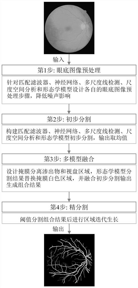
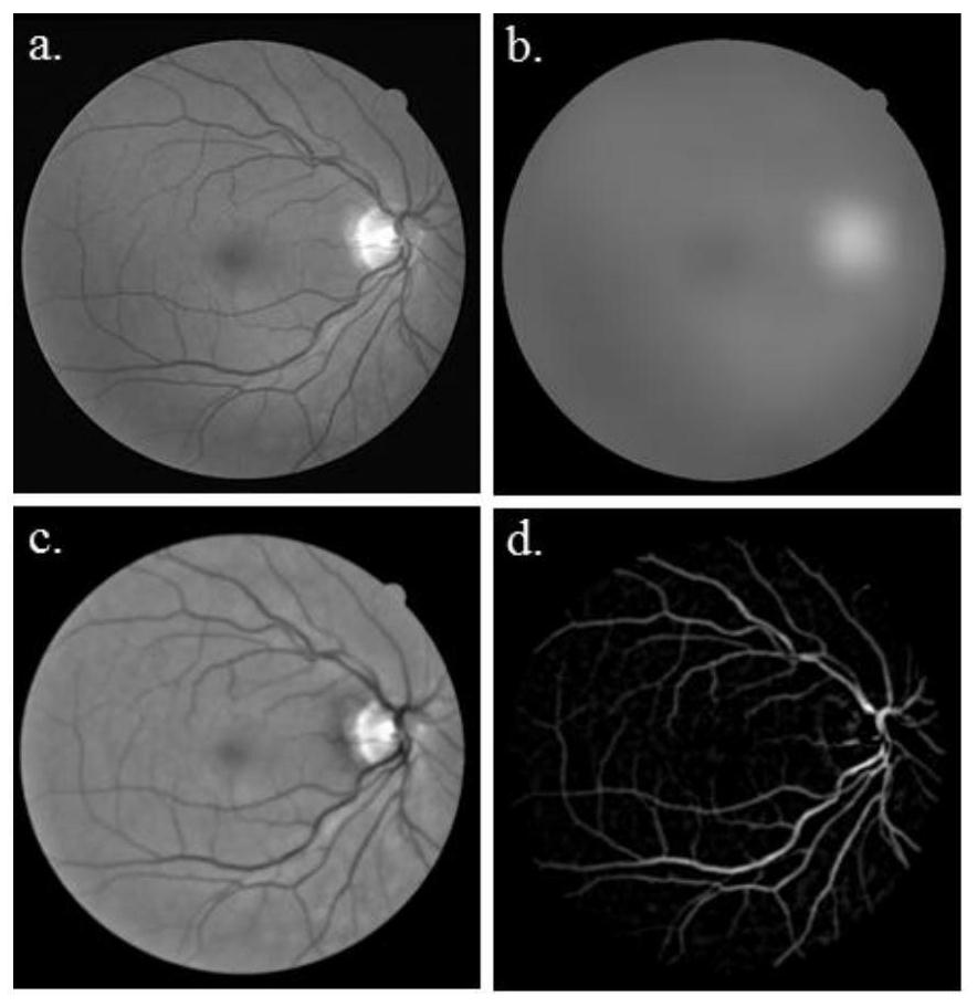
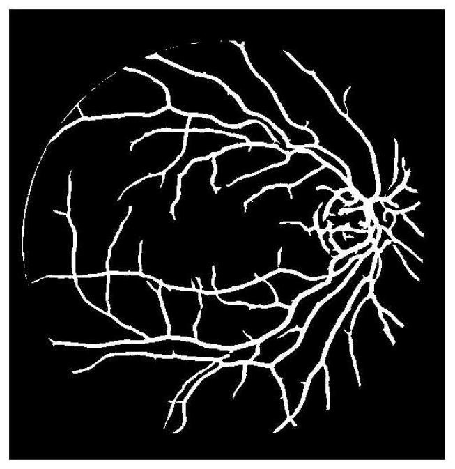
[0131] Embodiment 1, the retinal blood vessel automatic segmentation method facing glaucoma clinical diagnosis, such as Figure 1-9 shown, including the following:

[0132] In the present invention, the fundus image is preprocessed first, and then the matching filter, neural network, multi-scale line detection, scale space analysis and morphological model are respectively constructed to initially segment retinal blood vessels, and the average value of the five segmentation results is taken as the preliminary segmentation output in order to reduce noise . Next, a mask was designed to separate the exudates and optic disc regions, the segmentation results of the morphological model were replaced with the white regions of the mask, and the preliminary segmentation outputs were fused to generate combined results. Finally, the prior knowledge of retinal vessels is considered (that is, the retinal vessel network is composed of vessel trees connected with vessel segments), and the fi...


PUM


Abstract

The present invention proposes an automatic retinal vessel segmentation method for clinical diagnosis of glaucoma. By fusing five models of matching filter, neural network, multi-scale line detection, scale-space analysis and morphology that depend on different image processing techniques, the optic disc is eliminated. and the effects of bright areas such as exudates. At the same time, the present invention does not require a large amount of data to establish a retinal vessel segmentation model, which greatly reduces the amount and complexity of data to be processed, is easy to implement, and can effectively improve the efficiency of retinal vessel segmentation. The present invention also uses the region growing method and gradient information to iteratively grow the background and blood vessel regions on the basis of the multimodal fusion results. The segmentation results have better continuity and smoothness, and can retain more details of retinal blood vessels and more Complete visual retinal vascular network, thereby effectively assisting ophthalmologists in diagnosing diseases and reducing the burden on ophthalmologists.

Description

technical field [0001] The invention relates to the fields of digital medical image processing and analysis and intelligent medical care, in particular to an automatic retinal blood vessel segmentation method for glaucoma clinical diagnosis. Background technique [0002] Glaucoma is the world's leading irreversible blinding eye disease, known as the "silent thief of sight". It is estimated that by 2020, the number of glaucoma patients worldwide will rise to 79.6 million. At present, the prevalence of open-angle glaucoma in people over 40 years old in my country has reached 2.6%, accounting for 2 / 3 of the total number of glaucoma patients, and the blindness rate is 15% to 30%, which is much higher than the average level of 8% in economically developed countries. Early detection, early diagnosis, and early treatment are very important to inhibit the development of glaucoma. Color fundus images can directly observe retinal vascular lesions and other lesions such as exudates a...

Claims


Application Information

Patent Timeline
Patent Type & Authority Patents(China)
IPC IPC(8): G06T7/11G06T7/136G06T7/155
CPCG06T2207/20081G06T2207/20084G06T2207/30041G06T2207/30101G06T7/11G06T7/136G06T7/155
Inventor 赖小波徐小媚金波刘玉凤吕莉莉
Owner ZHEJIANG CHINESE MEDICAL UNIVERSITY
Why Eureka
Try Eureka