Lung CT image computer aided system and method based on cluster analysis
A computer-aided, CT image technology, applied in the field of medical image analysis, can solve the problems of increased workload of doctors, misdiagnosis and missed diagnosis, and achieve the effect of avoiding the influence of human subjective factors, reducing the amount of calculation and time, and increasing the accuracy.
- Summary
- Abstract
- Description
- Claims
- Application Information
AI Technical Summary
Problems solved by technology
Method used
Image


Examples
Embodiment 1
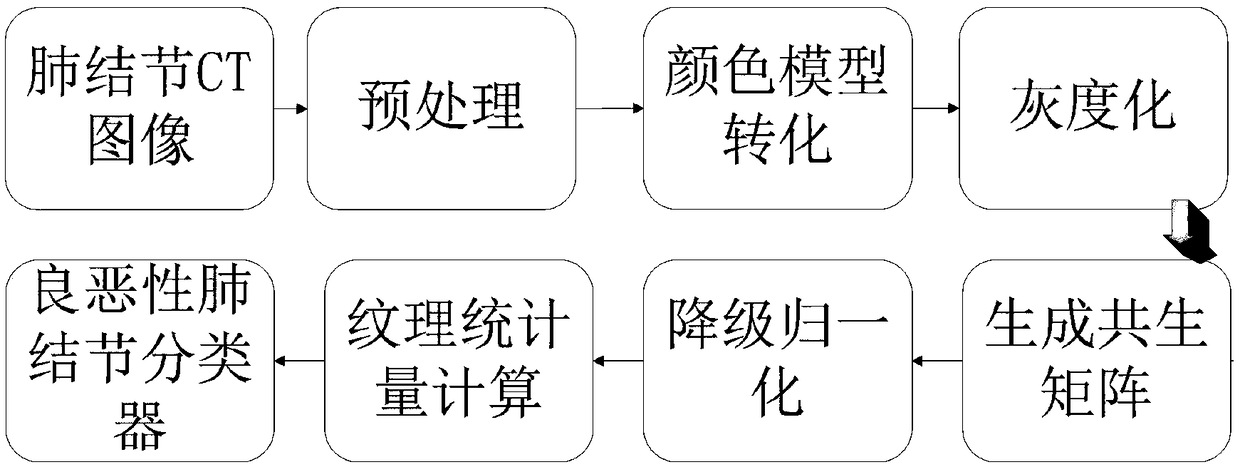
[0033] A method for feature extraction and classification of lung CT images based on cluster analysis, such as Figure 4 as shown,
[0034] Step 1, performing contour segmentation and extraction on CT images in advance, and marking benign nodules, and importing preprocessed images into the system for malignant nodules; figure 2 shown.
[0035] CT images of the lungs of patients were acquired by using CT scanning equipment. The CT scanner used Siemens Sensation 16-slice spiral CT to acquire CT plain scan cross-sectional images, and the image format was DICOM. The scanning parameters of CT equipment are tube voltage 120kV, tube current 220mAs, slice thickness 2-5mm, slice spacing 2-5mm, pitch 1-1.5, image reconstruction type B40, soft tissue display window, and cross-sectional image resolution 512 × 512 pixels, 10-15 cross-sectional images per patient. The method of the invention uses the patient's data for analysis and utilization, and then performs contour separation and e...
PUM
 Abstract
Description
Claims
Application Information