Specific marker Nagorno-Karabakh deformation microsporidia nucleic acid probe and fluorescence in-situ hybridization detection method thereof

A fluorescence in situ hybridization and nucleic acid probe technology, which is applied in the field of in situ hybridization detection of N. naga proteus in silkworm parasitic cyst tissue, can solve the problem that the FISH labeling method of N. naga proteus has not been invented yet, and the structure cannot be cultured in vitro , Weak research foundation and other issues, to achieve the effect of intuitive and reliable results, easy operation, simple and convenient operation

Inactive Publication Date: 2018-12-25
SOUTHWEST UNIVERSITY
View PDF1 Cites 0 Cited by
  • Summary
  • Abstract
  • Description
  • Claims
  • Application Information

AI Technical Summary

Problems solved by technology

However, due to the weak preliminary research foundation of N. naga proteus, lack of sufficient molecular data, and its special structure, which cannot be cultured in vitro, the FISH labeling method of N. naga proteus has not yet been invented.

Method used


Image

  • Specific marker Nagorno-Karabakh deformation microsporidia nucleic acid probe and fluorescence in-situ hybridization detection method thereof
  • Specific marker Nagorno-Karabakh deformation microsporidia nucleic acid probe and fluorescence in-situ hybridization detection method thereof
  • Specific marker Nagorno-Karabakh deformation microsporidia nucleic acid probe and fluorescence in-situ hybridization detection method thereof

Examples


Embodiment 1

[0032] Embodiment 1, probe design

[0033] According to the DNA sequence of VnBM ribosomal RNA large subunit (LSU rRNA) and its secondary structure model, combining with FISH probes requires the design and screening of FISH probes. The requirements for the FISH probe are: the length is about 18nt, and the Tm value is 48-60°C. If the Tm value is greater than 60°C or less than 48°C, the length of the probe can be adjusted appropriately. According to the above principles, a nucleic acid probe was designed, the sequence of which is as follows:

[0034] VnLSU-V1: 5'-gtattctattacgaccttc-3' (SEQ ID NO.1);

[0035] The nucleic acid probe designed by the technical solution can specifically bind the ribosomal RNA outside the nucleus, while the general probe can only bind the DNA in the nucleus and cannot achieve the purpose of distinguishing the microsporidia of silkworm in different stages.

[0036] The Naga proteus nucleic acid probe is coupled with different fluorescent markers as ...

Embodiment 2

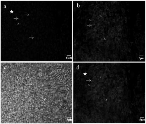
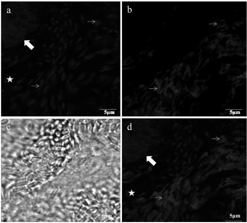
[0039] Embodiment 2, the method and result of VnLSU-V1 in situ hybridization experiment

[0040]1) Preparation of silkworm parasitic bursa tissue infected with VnBM

[0041] Bombyx mori were reared to the third instar, and the Naka proteus was evenly spread on the mulberry leaves, and 10 5 The amount of spores / silkworms was added to feed silkworms, and the growth and pathological changes of silkworms were observed and recorded. After the silkworm enters the fifth instar, the silkworm is dissected and the material obtained is the parasitic sac formed in the muscle cells of the rear midgut of the silkworm. Use tweezers to tear off the white tumor-like parasitic sac particles in the rear part of the silkworm midgut and place them in 1.5mL centrifuge tubes, take 6 tubes, fix 3 tubes with 4% paraformaldehyde, fix 3 tubes with 2.5% glutaraldehyde, and paste Label and store at 4°C.

[0042] 2) FISH probe labeling of hybridized VnBM in silkworm parasitic bursa

[0043] Put the par...

Embodiment 3

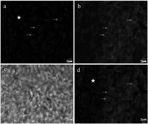
[0051] Embodiment 3, the method and result of VnLSU-V1-Cy3 in situ hybridization experiment

[0052] 1) Preparation of silkworm parasitic bursa tissue infected with VnBM

[0053] Bombyx mori were reared to the third instar, and the Naka proteus was evenly spread on the mulberry leaves, and 10 5 The amount of spores / silkworms was added to feed silkworms, and the growth and pathological changes of silkworms were observed and recorded. After the silkworm enters the fifth instar, the silkworm is dissected and the material obtained is the parasitic sac formed in the muscle cells of the rear midgut of the silkworm. Use tweezers to tear off the white tumor-like parasitic sac particles in the rear part of the silkworm midgut and place them in 1.5mL centrifuge tubes, take 6 tubes, fix 3 tubes with 4% paraformaldehyde, fix 3 tubes with 2.5% glutaraldehyde, and paste Label and store at 4°C.

[0054] 2) FISH probe labeling of hybridized VnBM in silkworm parasitic bursa

[0055] Put th...


PUM


Abstract

The invention relates to a specific marker Nagorno-Karabakh deformation microsporidia nucleic acid probe and a fluorescence in-situ hybridization detection method thereof. The nucleotide sequence of the nucleic acid probe is as shown in SEQ ID NO. 1, and in-situ hybridization particularly includes the steps: fixing and then decolorizing silkworm parasitism sac tissues infected by Nagorno-Karabakhdeformation microsporidia; adding hybridization solution containing the nucleic acid probe for hybridization and cleaning the silkworm parasitism sac tissues after hybridization; marking cell nucleuses of parasitism sacs and cell nucleuses of the Nagorno-Karabakh deformation microsporidia by DAPI, and observing fluorescent probe marking results by a fluorescent microscope. The nucleic acid probe is high in combination sensitivity to the Nagorno-Karabakh deformation microsporidia, higher in specificity and simple and convenient to operate, results are more visual and reliable, and pathogens ofall development stages except for mature spores in the parasitism sacs can be marked.

Description

technical field [0001] The invention belongs to the field of biomolecular detection, and relates to a nucleic acid probe for specifically marking Microsporidium nagaproteus, and also relates to a method for using the probe to detect Microsporidium nagaproteus in parasitic cyst tissue of silkworm by fluorescence in situ hybridization. Background technique [0002] Vairimorpha necatrix BM (VnBM) is an important pathogen of silkworm. In silkworms infected with VnBM, the intestinal tissue became swollen, the color changed to yellowish brown, and a large number of white tumor-like parasitic sacs were formed in the posterior part of the midgut. Because VnBM is an obligate intracellular parasite, it cannot be cultured in vitro, and its spore structure is special, so most conventional research methods cannot be used or have not yet been established. Important features such as the pathogenesis are still unclear. [0003] Currently, there are three commonly used observation methods ...

Claims


Application Information

Patent Timeline
Patent Type & Authority Applications(China)
IPC IPC(8): C12Q1/6888C12Q1/6841C12N15/11
CPCC12Q1/6841C12Q1/6888C12Q2563/107Y02A50/30
Inventor 李田房卓亚罗堿徐金智周泽扬
Owner SOUTHWEST UNIVERSITY
Why Eureka
Try Eureka