Carotid bifurcation three-dimensional cell culture model for drug screening, and construction method thereof

A three-dimensional cell, carotid artery technology, applied in drug screening, artificial cell constructs, animal cells, etc., can solve the problem that phenotype and function cannot reflect endothelial cell function well, and achieve accurate evaluation of cell function.

Inactive Publication Date: 2019-02-01
XI AN JIAOTONG UNIV
View PDF4 Cites 0 Cited by
  • Summary
  • Abstract
  • Description
  • Claims
  • Application Information

AI Technical Summary

Problems solved by technology

In previous in vitro experiments, endothelial cells were cultured on two-dimensional TCP. Due to the difference between the culture environment and the body, the phenotype and function of the cells may change, which may not reflect the function of endothelial cells in vivo.

Method used


Image

  • Carotid bifurcation three-dimensional cell culture model for drug screening, and construction method thereof
  • Carotid bifurcation three-dimensional cell culture model for drug screening, and construction method thereof
  • Carotid bifurcation three-dimensional cell culture model for drug screening, and construction method thereof

Examples


Embodiment 1

[0044] 1. Using HUVEC ( CRL-1730TM) cells were cultured, and the cells were routinely cultured with DMEM (Low Glucose), digested with trypsin and centrifuged at a low speed of 800-1000rpm for 5-10min, adding an appropriate amount of medium containing 8-12% fetal bovine serum to obtain a concentration of 1×10 5 ~2×10 6 Cells / ml of HUVEC suspension.

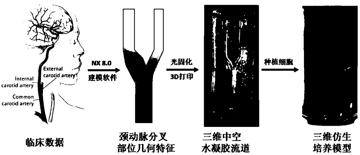
[0045] 2. Pour the cell suspension into the prepared hydrogel bionic blood vessel, and form a cell layer on the inner surface of the hydrogel bionic blood vessel structure to form a bionic blood vessel. Add the medium into the flow channel so that the medium can fully contact the HUVEC, and incubate in a gas incubator at 37°C, 5% carbon dioxide, and 95% air, such as figure 1 shown.

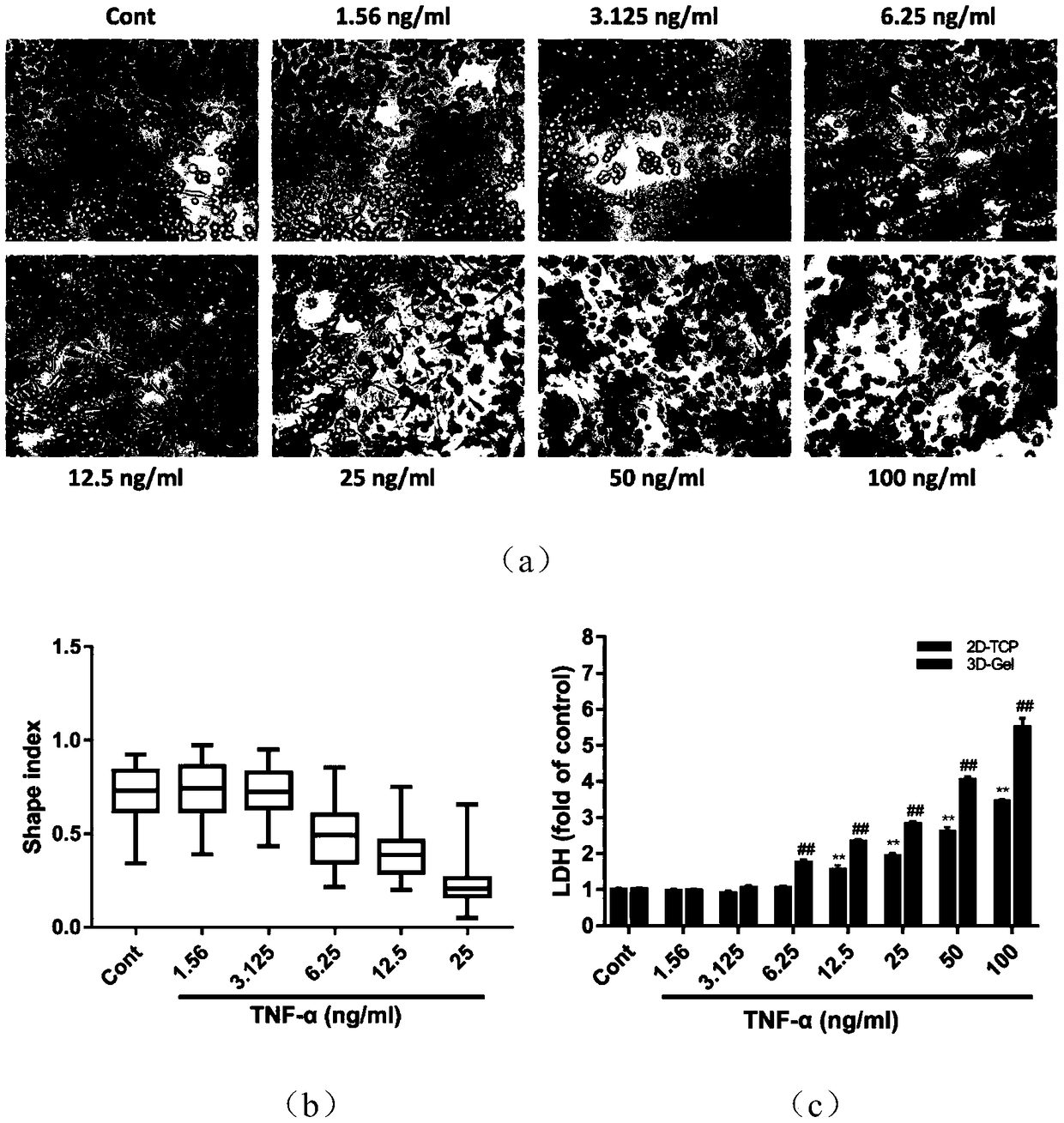
[0046] 3. See figure 2 , when the cells adhere to the wall and grow completely on the inner surface, such as figure 2 As shown in (a) Cont group, the endothelial cells grow evenly, the cells are oval, and the growth state is good. After the ...

Embodiment 2

[0049] 1. Add 1.56, 3.125, 6.25, 12.5, 25, 50, 100 ng / ml TNF-α culture solution into the model, after 24 hours of stimulation, observe the morphology under light microscope.

[0050] The result is as figure 2 As shown in middle (a), in the normal control group, the endothelial cells grow evenly, the cells are oval, and the growth state is good; the morphology of the endothelial cells begins to change significantly under the stimulation of 6.25ng / ml TNF-α, and the cells change from oval to The deformation changed to elongated, and the shape index decreased significantly and changed in a dose-dependent manner; under the stimulation of 50ng / ml TNF-α, some cells began to break and disintegrate. When the dose of TNF-α was 100ng / ml, the cells died in large numbers, and only cell fragments remained ;

[0051] 2. Use kits to detect LDH secreted by endothelial cells to reflect the degree of endothelial cell membrane damage.

[0052] from figure 2 In (c), it can be seen that in the...


PUM


Abstract

The invention discloses a carotid bifurcation three-dimensional cell culture model for drug screening, and a construction method thereof. Human-derived cells are used to make up for the defects of different species of animal models, and are closer to the real microenvironment of human bodies than traditional two-dimensional planar environments. The method can reflect the conditional controllability and intuitiveness of the traditional 2D cell culture, and can link a 2D cell culture system with tissue and organ researches to effectively simulate the influences of different pathological stimulion in-vivo cell functions, so the evaluation of the cell functions is accurate; and compared with existing three-dimensional vascular structure culture models, the model constructed by the method cangive stimulating factors in different pathological environments of simulated human bodies in order to study the changes of the form and functions of the cells in a disease state.

Description

technical field [0001] The invention belongs to the technical field of cell model preparation, and relates to a carotid bifurcation three-dimensional cell culture model for drug screening and a construction method thereof. Background technique [0002] According to the statistics of the World Health Organization, cardiovascular disease is the number one cause of death in the world. By 2030, the number of people dying of cardiovascular disease will increase to 23.3 million, and the cost of its treatment may be as high as 20 trillion yuan. The prevalence of cardiovascular disease risk factors in China is obvious, leading to a continuous increase in the number of cardiovascular disease incidence. The number of patients with cardiovascular disease will continue to increase rapidly in the next 10 years. At present, the death of cardiovascular disease accounts for the first place in the total death of urban and rural residents, 45.01% in rural areas and 42.61% in cities. The dis...

Claims


Application Information

Patent Timeline
IPC IPC(8): C12N5/071
CPCC12N5/069C12N2503/02C12N2513/00
Inventor 林蓉王博陈若梦刘亚雄王维蓉杨广德何延浩商晨旭姚凤高鸿倩
Owner XI AN JIAOTONG UNIV
Why Eureka
Try Eureka