Method for separating single blastomere in blastula

A separation method and a technology of blastomeres, which are applied in cell dissociation methods, biochemical equipment and methods, embryonic cells, etc., can solve the problems of time-consuming operators and expensive instruments and equipment, and achieve simple and easy steps and excellent cell morphology. full effect

Pending Publication Date: 2019-04-12
ANHUI AGRICULTURAL UNIVERSITY
View PDF5 Cites 0 Cited by
  • Summary
  • Abstract
  • Description
  • Claims
  • Application Information

AI Technical Summary

Problems solved by technology

The existing single cell separation technologies mainly include serial dilution method, micromanipulation method, fluorescence flow sorting method, laser capture microdissection technology and mic

Method used


Image

  • Method for separating single blastomere in blastula
  • Method for separating single blastomere in blastula

Examples


Embodiment 1

[0040] A method for separating a single blastomere in a blastocyst, comprising the steps of:

[0041] 1.1 Preparation of mouth aspiration needle

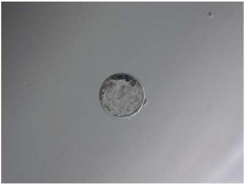
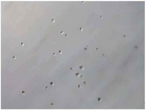
[0042] Take a BJ-40 thin glass tube and draw it into a mouth suction needle with a diameter of 145-155μm, a thin section length of 9-12cm, and a uniform thickness. Put the front part of the thin section of the mouth suction needle on an alcohol lamp and burn it After pulling it apart, the front part of the needle will become thinner after pulling it apart. At this time, use a grinding wheel to cut off the first 3 / 4 part of the thinner needle section. Note that the cut should be flat. Slightly burn the outside of the blue flame to make the needle mouth round.

[0043] 1.2 Preparation of separation needle

[0044] Take a BJ-40 thin glass tube and draw it into a mouth suction needle with a diameter of 145-155μm, a thin section length of 9-12cm, and a uniform thickness. Put the front part of the thin section of the mouth suction needl...


PUM


Abstract

The invention discloses a method for separating a single blastomere in a blastula. The method comprises the following steps: preparation of a mouth sucking needle; preparation of a separating needle;preparation of pronase and trypsin; removal of a transparent band of the blastula; and separation of the single blastomere: incubating a contracted blastula in 50 [mu]L of trypsin droplets for 40 min,transferring the blastula to 1 g/L of BSA droplets after incubation by using the narrow mouth sucking needle, conducting washing, rapidly blowing the blastula by using the narrow mouth sucking needleuntil some cell clusters cannot be separated by blow and suction, and then separating the individual cells in the cell cluster by a fine segment of the separating needle. The invention adopts the trypsin to separate the single blastomere in the blastula, the cell morphology is complete, and separation among the cells is clear. The reagents needed for invention are easy to obtain and economical; the steps are simple and easy; and therefore, the simple, cost-effective and practical method can be widely promoted.

Description

technical field [0001] The invention belongs to the field of cell biology, in particular to a method for separating a single blastomere in a blastocyst. Background technique [0002] With the development of reproductive medicine, more and more infertile couples choose assisted reproductive technology to conceive. The quality evaluation of human embryos generally only starts from the two perspectives of shape and development speed, but this cannot completely predict the embryo quality. Therefore, there are still some low-quality embryos that can further develop into expanded blastocysts. Previous studies have shown that frozen embryos with less than 50% blastomere survival rate can also achieve pregnancy, and even frozen embryos that are severely damaged during resuscitation can continue to divide and implant, which fully demonstrates that a single cleavage Balls have the ability to develop into blastocysts. The developmental ability of a single blastomere reflects the deve...

Claims


Application Information

Patent Timeline
IPC IPC(8): C12N5/073
CPCC12N5/0604C12N2509/00
Inventor 曹祖兵宁伟程竹兵刘洪瑜王恒刘传江高迪许腾腾张运海
Owner ANHUI AGRICULTURAL UNIVERSITY
Why Eureka
Try Eureka