Extracting method of extracellular vesicles and kit

An extraction method and kit technology, applied in the field of biological separation and extraction, can solve the problems of unfavorable separation scale, amplification, and high cost of experimental reagents, and achieve the effects of maintaining good biological activity of samples, improving processing efficiency, and low cost of use

Pending Publication Date: 2019-05-31
易春 +2
View PDF3 Cites 12 Cited by
  • Summary
  • Abstract
  • Description
  • Claims
  • Application Information

AI Technical Summary

Problems solved by technology

[0010] The fourth is the immunoaffinity method. This method has high specificity, simple operation, and does not require expensive equipment. However, since no universal marker for EVs has been found, the specific antibodies generally used can recognize EVs subgroups. The cost of reagents is relatively high, and it is not conducive to the scale-up of separation
This patent still uses centrifugal extraction of polymer precipitation, and inevitably there are problems in the centrifugal extraction of polymer precipitation described above.

Method used


Image

  • Extracting method of extracellular vesicles and kit
  • Extracting method of extracellular vesicles and kit
  • Extracting method of extracellular vesicles and kit

Examples


Embodiment 1

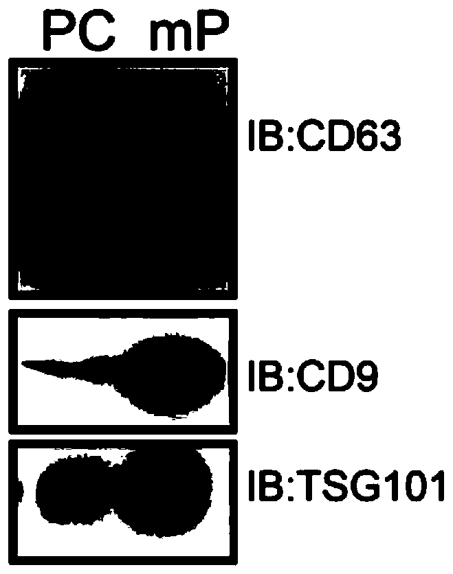
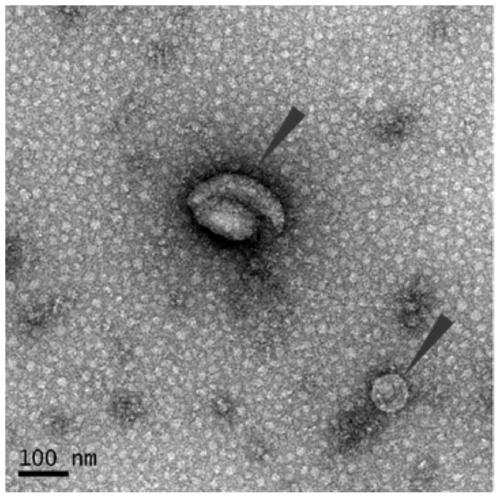
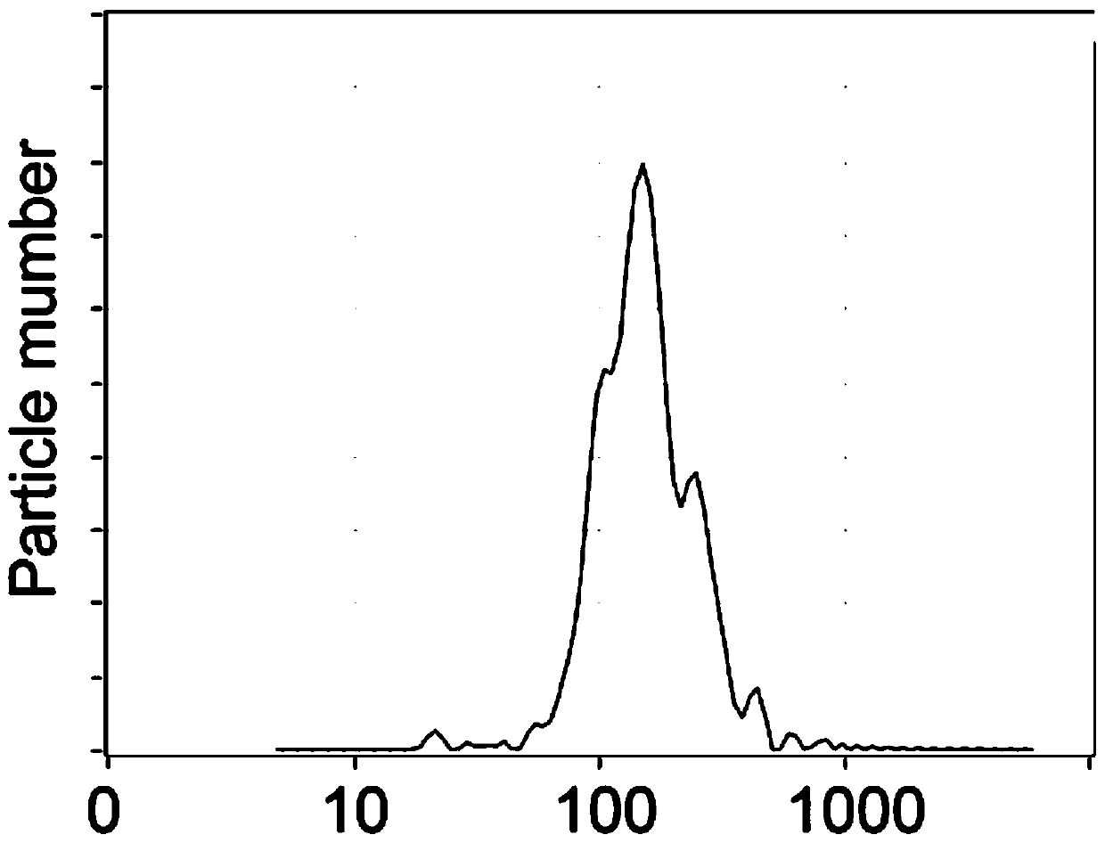
[0074] A method for purifying exosomes derived from mouse blood, the steps are as follows (specific results combined with Figure 1-Figure 3 ):

[0075] (1) Liquid preparation for exosome purification

[0076] a. Prepare equilibrium buffer solution (PBS, pH7.2), vacuum filter with 0.025 μm mixed cellulose ester (MCE), and equilibrate to 22±2°C for later use;

[0077] b. Prepare washing buffer (2M NaCl, 1M NaOH, 30% isopropanol), vacuum filter 0.025 μm mixed cellulose ester (MCE), and equilibrate to 22±2°C for later use;

[0078] d.ddH 2 O 0.025μm mixed cellulose ester (MCE) vacuum filtration, equilibrate to 22±2℃ for later use;

[0079] e. Prepare preservation solution with 20% ethanol, vacuum filter with 0.1 μm mixed cellulose ester (MCE), and balance to 22±2°C for later use;

[0080] (2) Mouse plasma preparation

[0081] a. Under sterile conditions, use a 3.2% sodium citrate anticoagulant vacuum blood collection tube of 3ml size to collect blood from 8-week-old male SPF...

Embodiment 2

[0149] A method for purifying exosomes derived from human urine, the steps are as follows (specific results combined with Figure 4-Figure 6 ):

[0150] (1) Liquid preparation for exosome purification

[0151] a. Prepare equilibrium buffer (PBS, pH 6.0), vacuum filter 0.025 μm mixed cellulose ester (MCE), and equilibrate to 22±2°C for later use;

[0152] b. Prepare washing buffer (2M NaCl, 1M NaOH, 30% isopropanol), vacuum filter 0.025 μm mixed cellulose ester (MCE), and equilibrate to 22±2°C for later use;

[0153] d.ddH2O 0.025μm mixed cellulose ester (MCE) vacuum filtration, balance to 22±2℃ for later use;

[0154] e. Prepare preservation solution with 20% ethanol, vacuum filter with 0.1 μm mixed cellulose ester (MCE), and balance to 22±2°C for later use;

[0155] (2) Collection of mid-morning urine

[0156] a. Under normal conditions, use a urine cup with a cover to collect about 100ml of mid-morning urine;

[0157] b. Within 2-3 minutes after collection, pour the uri...

Embodiment 3

[0230] A method for purifying Escherichia coli outer membrane vesicles, specifically as follows (the specific results are combined with Figure 7-Figure 8 ):

[0231] (1) Liquid preparation for purification of Escherichia coli outer membrane vesicles

[0232] a. Prepare equilibrium buffer (Tris-HCl, pH8), vacuum filter 0.025 μm mixed cellulose ester (MCE), and equilibrate to 22±2°C for later use;

[0233] b. Prepare washing buffer (2M NaCl, 0.5M NaOH, 30% isopropanol), vacuum filter 0.025 μm mixed cellulose ester (MCE), and equilibrate to 22±2°C for later use;

[0234] d.ddH2O 0.025μm mixed cellulose ester (MCE) vacuum filtration, balance to 22±2℃ for later use;

[0235] e. Prepare preservation solution with 20% ethanol, vacuum filter with 0.1 μm mixed cellulose ester (MCE), and balance to 22±2°C for later use;

[0236] (2) Escherichia coli culture supernatant collection

[0237] a. Autoclave steam sterilized bamboo toothpicks to pick a small amount of liquid nitrogen to p...


PUM

PropertyMeasurementUnit
particle sizeaaaaaaaaaa
pore sizeaaaaaaaaaa
heightaaaaaaaaaa

Abstract

The invention relates to the technical field of biological separation and extraction, in particular to an extracting method of extracellular vesicles. The method comprises the steps that a biologicalsample is provided; the biological sample is in contact with a capturing surface at least twice on the condition that impurities in the biological sample are partially or wholly reserved on the capturing surface, wherein the impurities are partial or whole other substances except the extracellular vesicles in the biological sample, and the capturing surface comprises an inner surface and/or outersurface of a spherical-particle porous material; when the impurities are partially or wholly reserved on the surface of a capture, breakthrough liquid is extracted liquid containing the extracellularvesicles; or the breakthrough liquid is further concentrated. By means of the method, the epicyte vesicles can be quickly separated and purified, the separation operation is simple, the single use cost is low, the separated sample in high in purity, the bioactivity of the sample is kept good, and the extracting method is applied to fundamental research in the aspects of biomarker discovery, biotherapy and the like and industrial production.

Description

technical field [0001] The invention relates to the technical field of biological separation and extraction, in particular to an extraction method and kit for extracellular vesicles. Background technique [0002] Extracellular vesicles (EVs) are a class of membrane-containing vesicles released from prokaryotes to higher eukaryotes and plant cells through an evolutionarily conserved pathway. Over the past few decades, extracellular vesicles have been considered as the key to prokaryotic and eukaryotic cell-to-cell communication due to their ability to transport proteins, lipids, nucleic acids, and sugars to affect different physiological and pathological functions of target and generating cells. important carrier. Based on size, extracellular vesicles can be divided into small extracellular vesicles (<300nm, small EVs, sEVs) and medium and large extracellular vesicles >300nm (medium / large EVs, m / lEVs) [0003] Exosomes (exosomes) are a subclass of extracellular vesicl...

Claims


Application Information

Patent Timeline
Patent Type & Authority Applications(China)
IPC IPC(8): C12N5/078C12N5/071C12N1/20C12R1/19
Inventor 陆路
Owner 易春
Why Eureka
Try Eureka