Application of tanshinone IIA in inhibiting abnormal accumulation of Tau protein

A tanshinone and protein technology, applied in the field of biomedicine, can solve problems such as unsatisfactory drug efficacy, achieve the effects of less toxic and side effects, good curative effect, and inhibiting abnormal aggregation of Tau protein

Inactive Publication Date: 2019-06-21
SHENZHEN UNIV
View PDF2 Cites 1 Cited by
  • Summary
  • Abstract
  • Description
  • Claims
  • Application Information

AI Technical Summary

Problems solved by technology

[0006] The purpose of the present invention is to overcome the above-mentioned deficiencies of the prior art, to provide a kind of application of tanshinone IIA as an inhibitor of abnormal aggregation of Tau protein or in a drug, so as to solve the clinical problems of existing drugs for inhibiting abnormal aggregation of Tau protein and treating tau protein diseases. Unsatisfactory technical problems

Method used


Image

  • Application of tanshinone IIA in inhibiting abnormal accumulation of Tau protein
  • Application of tanshinone IIA in inhibiting abnormal accumulation of Tau protein
  • Application of tanshinone IIA in inhibiting abnormal accumulation of Tau protein

Examples


Embodiment 1

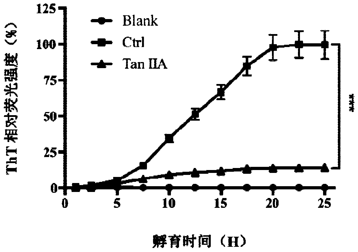
[0044] Example 1: Tanshinone IIA inhibits heparin-induced Tau protein aggregation

[0045] Tau protein can be induced by heparin sodium in vitro to self-aggregate to form fibers, thereby simulating the pathological process of abnormal aggregation of Tau protein in vivo to produce neurofibrillary tangles. Thioflavin T (ThT) can bind to the β-structure of fibers to emit fluorescence, so the fibrosis process of Tau protein can be tracked through the fluorescence value of ThT. Dissolve Tau protein to 20 μM with Tris-HCl buffer and add to black microplate, then add thioflavin T, heparin sodium and tanshinone IIA (20 μM) Under the excitation light of 485nm, continuous detection was carried out for 25 hours, and the relative fluorescence value was used to make a graph.

[0046] like figure 1 As shown, in vitro, Tau protein aggregates under the induction of heparin sodium, and when tanshinone IIA is added to the system, the aggregation of Tau protein is significantly inhibited.

Embodiment 2

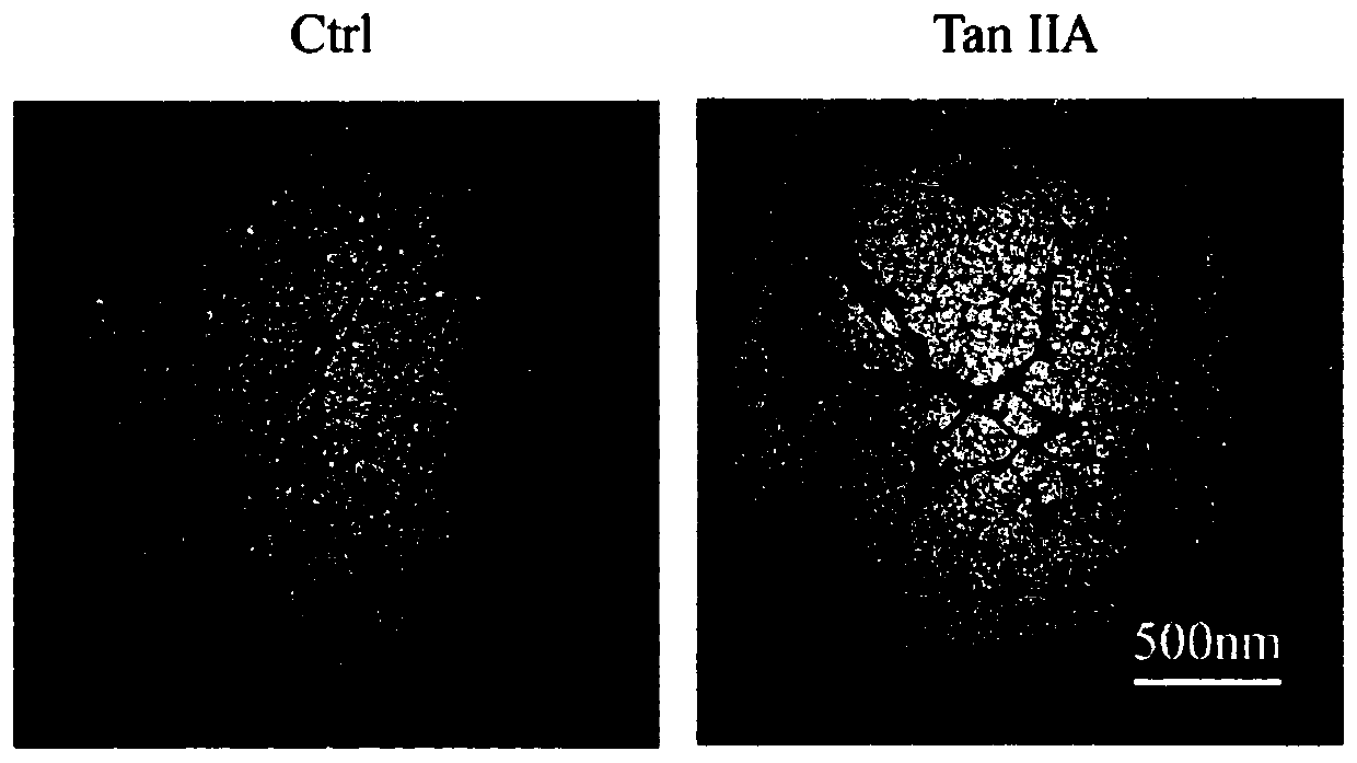
[0047] Example 2: Transmission Electron Microscopy Experiment of Tanshinone IIA Inhibiting Tau Protein Fibrosis

[0048] Methods: The fibrosis of Tau protein samples can be directly observed by transmission electron microscopy (TEM), which can further verify the results of ThT fluorescence experiments. Take 5 μL of each sample in the Tau protein ThT experiment, drop it on a 230-mesh copper grid, then absorb the excess sample with filter paper, drop 5 μL of uranyl acetate on each after drying, absorb the excess liquid after staining for 1 minute, dry and place in In a transmission electron microscope (NIPPON TEKNO, JEM-1230), observe and take pictures. Result: results like figure 2 As shown, dense Tau protein fibers were observed under the transmission electron microscope in the control group, but after the addition of tanshinone IIA, the Tau protein fibers were sparse and fractured, indicating that tanshinone IIA can effectively hinder the formation of Tau protein fibers.

Embodiment 3

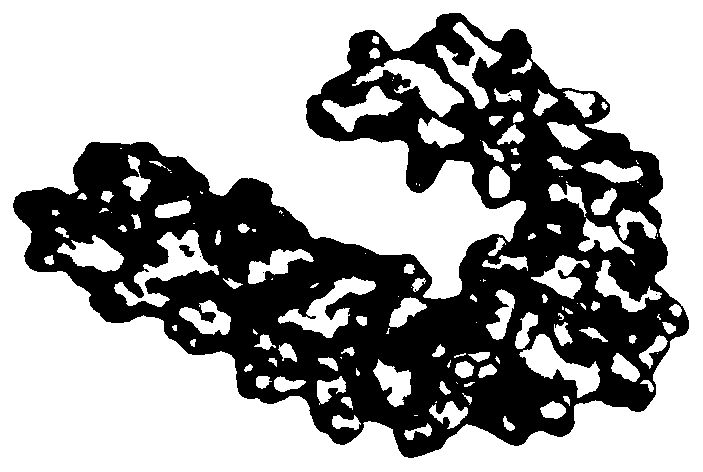
[0049] Example 3: Molecular docking of Tanshinone IIA and Tau protein

[0050] The Autodock vina 1.1.2 program was used for molecular docking to study the binding mode between tanshinone IIA and Tau protein. The core structure of the Tau protein fiber (PDB number 5O3L) was downloaded from the RCSB protein database (http: / / www.rcsb.org / pdb / home / home.do). The 2D results of Tanshinone IIA were drawn by ChemBioDraw Ultra 12.0 software, and the 3D structure of the molecule was optimized by ChemBio3D Ultra 12.0. Use AutoDockTools 1.5.6 to process and dock the protein after hydrogenation. The coordinates of the center point of the docking port are (215.348, 133.426, 155.072), size_x, size_y, and size_z are 15, 15, 15, and 15, respectively, and the global search exhaustiveness (exhaustiveness ) was set to 20, other parameters were set to default values, the best docking method was selected by docking scoring, and visual analysis was performed by PyMoL1.7.6 software.

[0051] like ...


PUM


Abstract

The invention discloses tanshinone IIA as an inhibitor of abnormal accumulation of Tau protein and application of tanshinone IIA in the preparation of drugs to prevent / treat Tau disease or in the application of functional foods or health products to prevent / relieve Tau protein disease. The tanshinone IIA discloses herein has the function of effectively inhibiting abnormal accumulation of Tau protein, thereby preventing pathological process of Tau protein disease; therefore, the tanshinone IIA can act as an anti-Tau protein disease drug to treat neurodegenerative diseases.

Description

technical field [0001] The invention belongs to the technical field of biomedicine, and in particular relates to the application of tanshinone IIA in a drug for inhibiting abnormal aggregation of tau protein and preventing / treating tau protein disease. Background technique [0002] In recent years, the number of patients with neurodegenerative diseases has increased year by year, manifesting as cognitive impairment, memory loss, and weakened behavior and motor abilities. Among them, neurofibrillary tangles in nerve cells are a common pathological feature. The tangles are formed by abnormal aggregation of tau protein, which leads to neuronal structural changes, functional degradation, and even apoptosis. Tau protein is a microtubule-related protein encoded by the MAPT gene. Under physiological conditions, it is abundantly present in neurons as a soluble phosphorylated protein, which induces and promotes the formation of microtubules, maintains the stability of microtubules, a...

Claims


Application Information

Patent Timeline
IPC IPC(8): A61K31/58A61P25/28A61P25/16A61P25/00A61P21/00
Inventor 续旭蔡楠陈家杰顾粮毕德成李梅婷杨朋
Owner SHENZHEN UNIV
Why Eureka
Try Eureka