

Adaptive multi-site scanning imaging method based on deep learning and system thereof
A scanning imaging and deep learning technology, applied in the field of nuclear medicine imaging, can solve problems such as lack of intuitive and clear connection, doubts about the clinical value of personalized collection procedures, complex optimization, etc., to achieve the effect of improving diagnostic efficiency
- Summary
- Abstract
- Description
- Claims
- Application Information
AI Technical Summary
Problems solved by technology
Method used
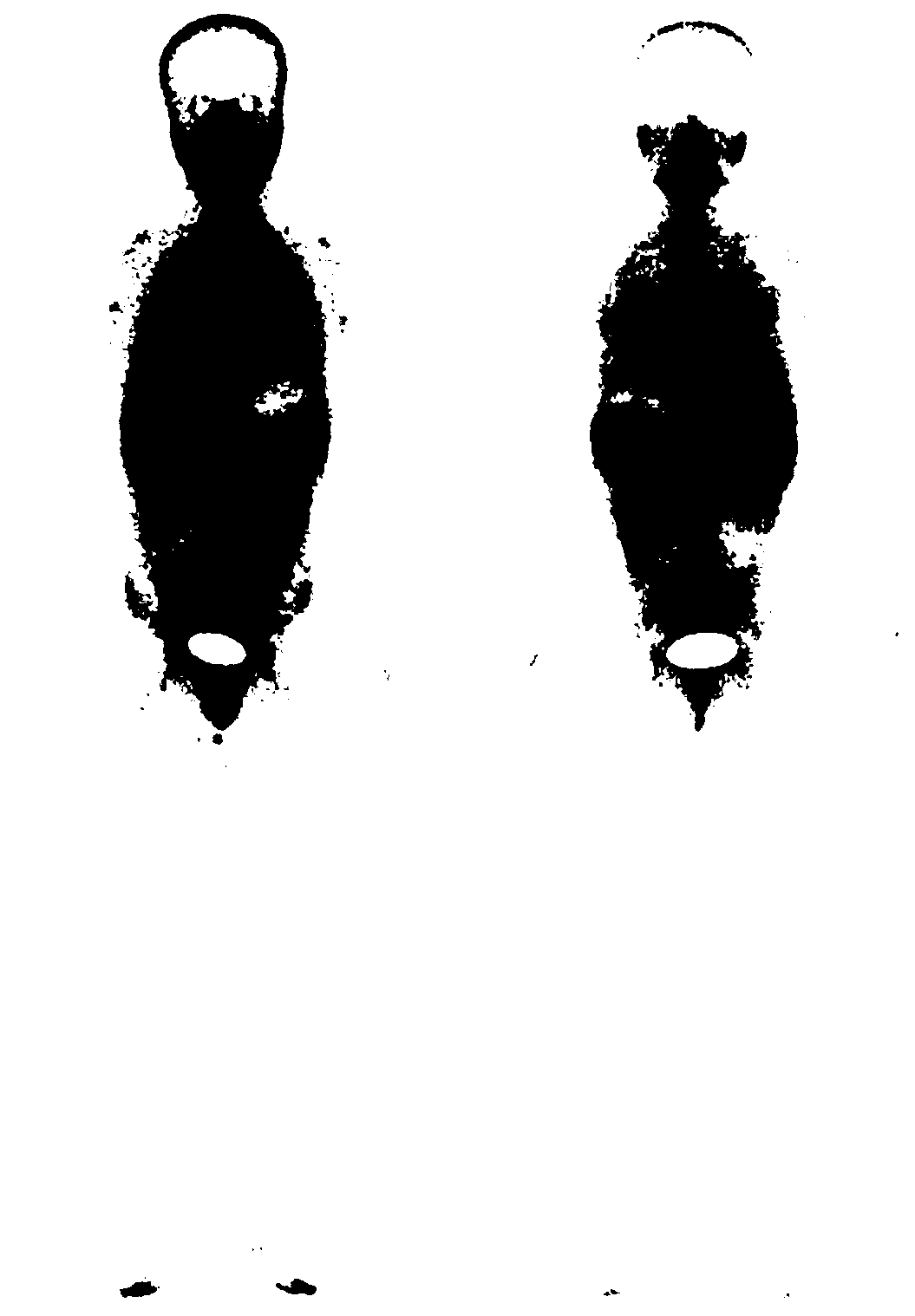
Image

Examples
Embodiment 1
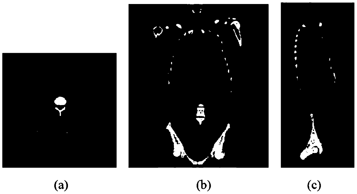
[0051] The adaptive multi-part scanning imaging method based on deep learning in this embodiment is applied to multi-modal imaging equipment mainly based on nuclear medicine, such as figure 1 shown, including the following steps:
[0052] Step A, performing single-modal or multi-modal reconnaissance scanning imaging for multiple target parts of the imaging target;
[0053] Step B, using the image analysis software based on deep learning technology to analyze the reconnaissance scanning image data in step A, and combining the relevant prior information of imaging target detection, detecting the local area that needs to be further focused on imaging, marking its boundary, and Quantitative evaluation of its importance or risk;
[0054] Step C, according to the detection and analysis results of step B, select the process and parameters to be optimized for the next scan and implement the scan.
[0055] Preferably, the nuclear medicine-based multimodal imaging equipment is a SPECT...
Embodiment 2
[0073] The adaptive multi-part scanning imaging system based on deep learning in this embodiment is applied to multi-modal imaging equipment mainly based on nuclear medicine, and the multi-modal imaging equipment based on nuclear medicine is embedded with technology based on deep learning. image analysis software and includes the following modules:
[0074] The reconnaissance scanning imaging module is used to perform single-modal or multi-modal reconnaissance scanning imaging for multiple target parts of the imaging target;
[0075] The quantitative evaluation module is used to analyze the reconnaissance scan image data generated by the reconnaissance scan imaging module by using image analysis software based on deep learning technology, and combine the relevant prior information of imaging target detection to detect local areas that need further key imaging, Mark its boundaries and quantify its importance or risks;
[0076] The selection optimization module is used to selec...
PUM
 Abstract
Description
Claims
Application Information