Chemiluminescence quantitative detection kit for myoglobin and preparation method and detection method thereof

A myoglobin and chemiluminescence technology, applied in the field of in vitro diagnostic reagents, can solve the problems of expensive reagents and instruments, limited coating density, long reaction time, etc., and achieve the effects of short detection time, reliable detection means and high sensitivity

Inactive Publication Date: 2019-07-09
DIRUI MEDICAL TECH CO LTD
View PDF1 Cites 11 Cited by
  • Summary
  • Abstract
  • Description
  • Claims
  • Application Information

AI Technical Summary

Problems solved by technology

There are following defects in the method for quantitatively detecting myoglobin above: 1) ELISA method adopts microporous plate as immobilization, and

Method used


Image

  • Chemiluminescence quantitative detection kit for myoglobin and preparation method and detection method thereof
  • Chemiluminescence quantitative detection kit for myoglobin and preparation method and detection method thereof
  • Chemiluminescence quantitative detection kit for myoglobin and preparation method and detection method thereof

Examples


Embodiment 1

[0056] Preparation of a chemiluminescence quantitative detection kit for myoglobin

[0057] 1) Streptavidin-coated magnetic particles

[0058] Take a certain amount of magnetic particles, wash 3 times with 20mM PB buffer, then dilute to 10mg / ml, add streptavidin according to the mass ratio of magnetic particles to streptavidin of 0.02%, and incubate at 37°C for 18h , placed on a magnetic separation rack to separate magnetic beads, removed the supernatant, washed 3 times with 20mM PB buffer, added blocking solution (20mM PB buffer containing 2% BSA), incubated at 37°C for 18h, and placed on a magnetic separation rack Separate the magnetic beads, remove the supernatant, wash 3 times with 20mM PB buffer, dilute to 10mg / ml with 20mM PB buffer and save for later use to obtain streptavidin-coated magnetic particles;

[0059] 2) Acridinium ester-labeled myoglobin antibody

[0060] Take a certain amount of myoglobin antibody, replace the myoglobin antibody preservation solution with...

Embodiment 2

[0064] Preparation of a chemiluminescence quantitative detection kit for myoglobin

[0065] 1) Streptavidin-coated magnetic particles

[0066] Take a certain amount of magnetic particles, wash 3 times with 20mM PB buffer, then dilute to 10mg / ml, add streptavidin according to the mass ratio of magnetic particles to streptavidin 1%, and incubate at 37°C for 24h , placed on a magnetic separation rack to separate magnetic beads, removed the supernatant, washed 3 times with 20mM PB buffer, added blocking solution (20mM PB buffer containing 5% BSA), incubated at 37°C for 24h, and placed on a magnetic separation rack Separate the magnetic beads, remove the supernatant, wash 3 times with 20mM PB buffer, dilute to 10mg / ml with 20mM MPB buffer and save for later use to obtain streptavidin-coated magnetic particles;

[0067] 2) Acridinium ester-labeled myoglobin antibody

[0068] Take a certain amount of myoglobin antibody, replace the myoglobin antibody preservation solution with 20mM...

Embodiment 3

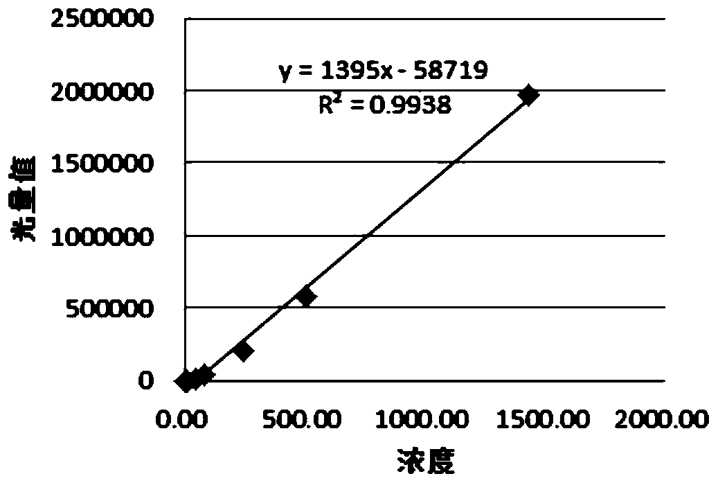
[0072] As a preferred scheme of a chemiluminescent quantitative detection kit for myoglobin in this embodiment, the mass ratio of the magnetic particle and streptavidin described in R1 is 0.072%; The molar ratio of acridinium ester is 1:3, and the concentration of acridinium ester-labeled myoglobin antibody is 0.4ug / ml. The molar ratio of myoglobin primary antibody to biotin described in R3 is 1:5, and the concentration of biotin-labeled myoglobin antibody is 1.4ug / ml;


PUM

PropertyMeasurementUnit
Linear correlation coefficientaaaaaaaaaa

Abstract

The invention discloses a chemiluminescence quantitative detection kit for myoglobin and a preparation method and a detection method thereof, belongs to the technical field of in vitro diagnostic reagents, and solves the technical problems of low sensitivity, long reaction time and high cost in the prior art. The kit comprises the following reagents: R: a buffer solution containing streptavidin coated magnetic particles; R2: a buffer solution containing a chemiluminescent marker-labeled myoglobin monoclonal antibody; and R3: a buffer solution containing coupling marker-labeled myoglobin monoclonal antibody. According to the chemiluminescence quantitative detection kit for the myoglobin provided by the invention, the principle of a double-antibody sandwich method is adopted, an avidin-coupling marker system is used, and chemiluminiscence is carried out by adopting a chemiluminescent marker, thus the detection sensitivity of the kit is greatly improved, the reaction time is shortened, the reagent cost is reduced, and the kit can be used for the quantitative detection of myoglobin contents in serum/plasma.

Description

technical field [0001] The invention relates to the technical field of in vitro diagnostic reagents, in particular to a chemiluminescence quantitative detection kit for myoglobin, a preparation method and a detection method thereof. Background technique [0002] The tertiary structure of sperm whale myoglobin was clarified by Kendrew in 1960 by X-ray diffraction method, which is the first protein tertiary structure described in the world. Myoglobin (MYO) is an intracellular oxyhemoglobin-binding protein ubiquitously present in cardiac and skeletal muscles with a molecular weight of 17,800 Daltons. In acute myocardial injury, MYO is first released into the blood. About 2-3 hours after the onset of symptoms, MYO in the blood can exceed the normal upper limit, reach the peak at 9-12 hours, and return to normal after 24-36 hours. Therefore, the determination of serum or plasma myoglobin can be used as the early and most sensitive index for the diagnosis of acute myocardial infa...

Claims


Application Information

Patent Timeline
IPC IPC(8): G01N33/68
CPCG01N33/6887G01N2333/805
Inventor 韩美玉王立英孙成艳高威何浩会
Owner DIRUI MEDICAL TECH CO LTD
Why Eureka
Try Eureka