Three-dimensional brain tumor image segmentation method based on improved U-Net neural network

An image segmentation and neural network technology, applied in the field of medical imaging, can solve the problems of low algorithm segmentation accuracy, category imbalance, small brain tumor image data set, etc., achieve high accuracy, prevent overfitting, and improve network performance.

Pending Publication Date: 2019-08-13
TIANJIN UNIV
View PDF3 Cites 66 Cited by
  • Summary
  • Abstract
  • Description
  • Claims
  • Application Information

AI Technical Summary

Problems solved by technology

[0007] In order to overcome the deficiencies of the existing technology, and aiming at the problems of small brain tumor image data sets, serious category imbalance, and low segmentation a...

Method used


Image

  • Three-dimensional brain tumor image segmentation method based on improved U-Net neural network
  • Three-dimensional brain tumor image segmentation method based on improved U-Net neural network
  • Three-dimensional brain tumor image segmentation method based on improved U-Net neural network

Examples


Embodiment Construction

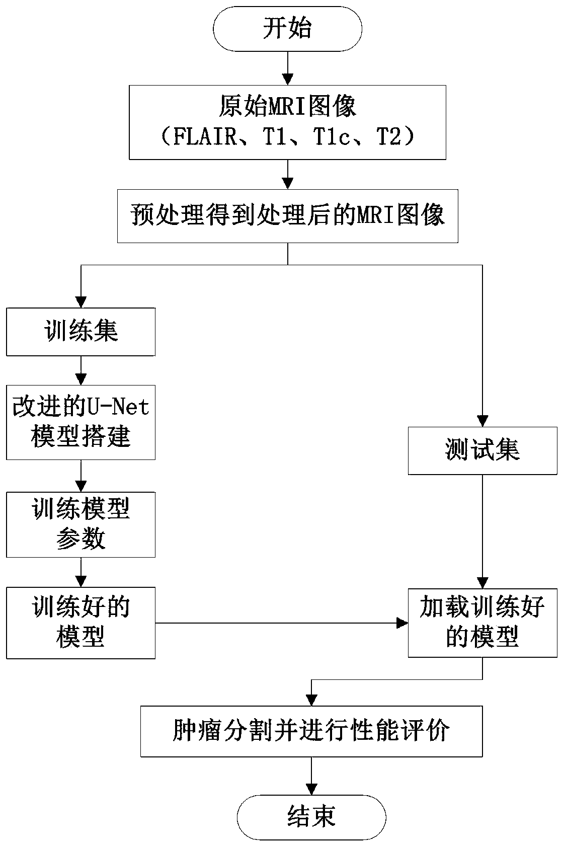
[0028] The invention combines medical images and deep learning algorithms to complete precise segmentation of three-dimensional brain tumor nuclear magnetic resonance images. Aiming at the problems of small brain tumor image data set, serious category imbalance, and low segmentation accuracy of existing algorithms, the present invention proposes a 3D brain tumor image segmentation method based on an improved U-Net convolutional neural network. figure 1 It is a block diagram of the algorithm proposed by the present invention. First, the four modes in the original MRI image are preprocessed respectively; secondly, the preprocessed images are divided into a training set and a test set, and an improved U-mode is built and trained on the training set. Net convolutional neural network model; finally, after the improved U-Net convolutional neural network model is trained, test the model on the test set, and use the corresponding evaluation indicators to evaluate the segmentation resul...


PUM


Abstract

The invention relates to a three-dimensional brain tumor image segmentation method based on an improved U-Net neural network. The method comprises the following steps: firstly, performing bias field effect removal processing of an N4ITK algorithm on three-dimensional brain tumor MRI image data, and secondly, respectively performing gray normalization preprocessing on each modal image in an original MRI image; building and training an improved U-Net convolutional neural network model; in a training process, inputting the four types of modal data of the patient as four channels of a neural network into an improved U-Net convolutional neural network model for training; the convolutional neural network U-Net being used as a basis, establishing an improved U-Net convolutional neural network model, and the improved U-Net convolutional neural network model comprising an analysis path used for extracting features and a synthesis path used for recovering a target object.

Description

technical field [0001] The invention is an important field in the field of medical imaging, and combines medical images with deep learning algorithms to complete precise segmentation of three-dimensional brain tumor nuclear magnetic resonance images. Background technique [0002] Intracranial tumors, also known as "brain tumors", are one of the most common diseases in neurosurgery. In terms of the incidence of systemic tumors, brain tumors rank fifth, only lower than stomach, uterus, breast and esophageal tumors. Magnetic Resonance Imaging (MRI) has unique advantages such as no radiation damage, no bony artifacts, multi-faceted multi-parameter imaging, and a high degree of soft tissue resolution. conditions for making a diagnosis and formulating a treatment plan. Due to the imaging principle and imaging conditions of medical instruments, as well as some other influencing factors, the obtained pictures will not be conducive to the observation of human eyes, so that doctors ...

Claims


Application Information

Patent Timeline
IPC IPC(8): G06T7/00G06T7/12
CPCG06T7/0012G06T7/12G06T2207/10088G06T2207/30016G06T2207/30096G06T2207/20081G06T2207/20084
Inventor 白柯鑫李锵关欣
Owner TIANJIN UNIV
Why Eureka
Try Eureka