Method for enriching or detecting astrocyte-derived exosomes from biological fluids

A technology of astrocytes and biological fluids, applied in the field of biomedicine, can solve the problems of routine application of traumatic operations, etc.

Inactive Publication Date: 2019-08-16
PEKING UNIV
View PDF2 Cites 4 Cited by
  • Summary
  • Abstract
  • Description
  • Claims
  • Application Information

AI Technical Summary

Problems solved by technology

[0004] The present invention aims to provide a method for enriching or detecting astrocyte-derived exosomes from biological fluids, so as to solve the existing biological problems for auxiliary

Method used


Image

  • Method for enriching or detecting astrocyte-derived exosomes from biological fluids
  • Method for enriching or detecting astrocyte-derived exosomes from biological fluids
  • Method for enriching or detecting astrocyte-derived exosomes from biological fluids

Examples


Embodiment approach

[0094] According to a typical embodiment of the present invention, the step (b) includes: (b) binding the astrocyte-derived exosomes in the biological fluid to a solid phase through an immune complex; (c) separating the solid phase binding exosomes and (d) determining the level of astrocyte-derived biomarkers in the solid-phase-bound exosomes isolated in step (c), and, compared with those from subjects without neurological disease Compared with the control level of , the difference in the level of biomarkers from astrocyte-derived exosomes in the subjects significantly indicates that the subjects suffer from neurological diseases.

[0095] In another typical embodiment of the present invention, when the biomarker is astrocyte-derived exosomes themselves, the anti-GLT1 antibody is a labeled antibody, and step (b) includes: directly detecting the astrocytes in the biological fluid by means of the label Stemocyte-derived exosomes; the labeling of anti-GLT1 antibodies includes but...

Embodiment 1

[0099] Exosome Isolation

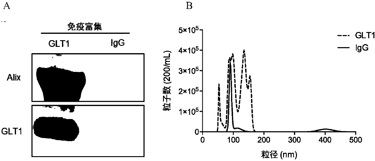
[0100] Exosomes were isolated from mouse or human plasma following a protocol modified from Tauro et al (Methods 56:293-304, 2012) using antibody-coated superparamagnetic microbeads.

[0101] Brief description: 10 μg of anti-GLT1 antibody (ab178401, Abcam, Cambridge, MA, USA), or normal mouse IgG (Santa Cruz Biotechnology, Dallas, TX, USA) (as a negative control) was coated on a group (1 mg) of M -270 epoxy beads using an antibody conjugation kit (Life Technologies, Grand Island, NY, USA) according to the manufacturer's instructions. After rapid thawing (within 2 minutes) at 37°C, plasma samples (>300 μL) were centrifuged at 2,000ⅹg for 15 minutes, followed by 12,000ⅹg for 30 minutes, and then washed with phosphate-buffered saline (PBS) (pH 7.4 ) to dilute the supernatant 1:3. One set of antibody-coated beads and 900 [mu]L diluted plasma were incubated for approximately 24 hours at 4[deg.] C. with gentle rotation. The beads were then washed four t...

Embodiment 2

[0105] Characterization of anti-GLT1-trapped exosomes from human plasma

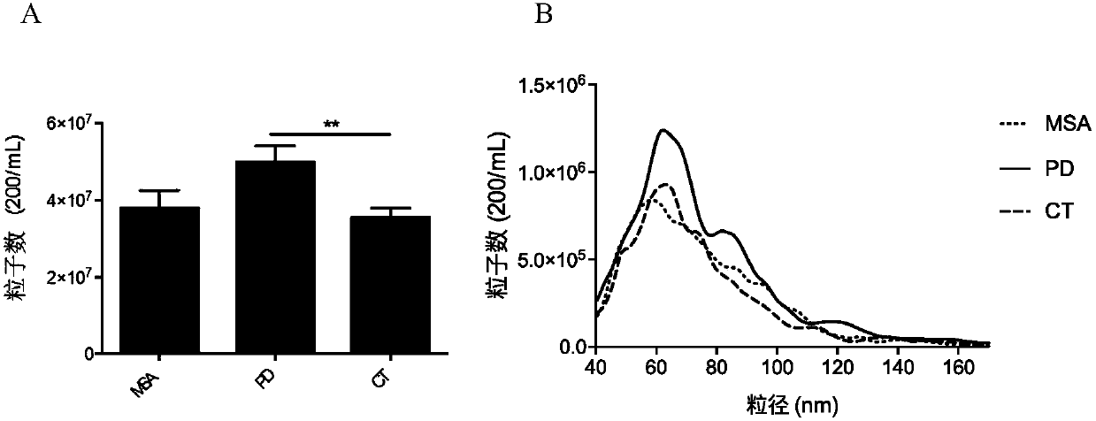
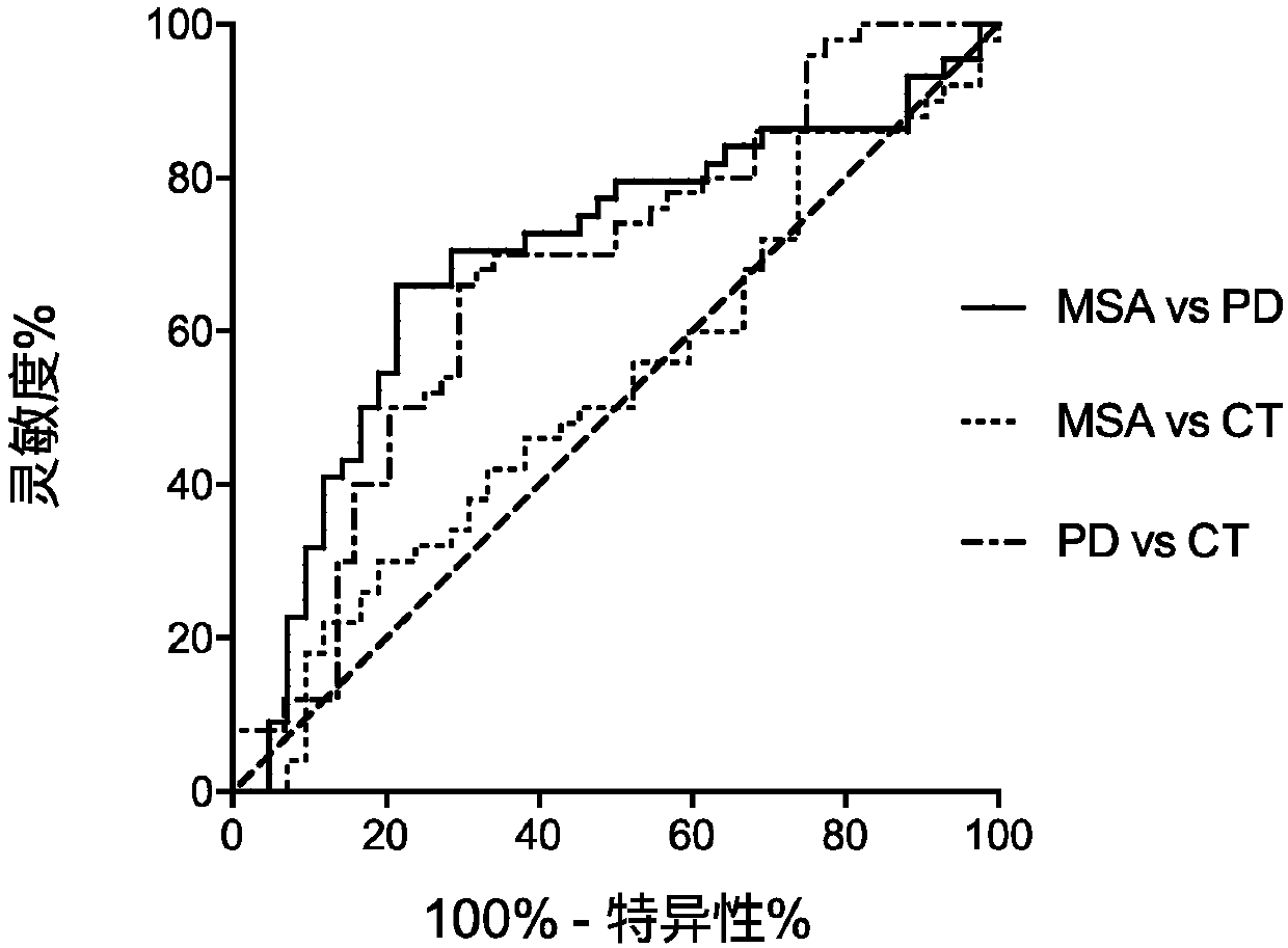
[0106] Luminex assay: α-syn, oligomer a-syn, PS129, tau, Aβ protein concentration.

[0107] Western Blot Analysis: Western blots were performed following standard protocols. Exosome samples (~10 μg protein) were lysed with Laemmli sample buffer and separated on 1D SDS-PAGE gels before transferring to polyvinylidene fluoride membranes. Proteins on the above membranes were detected with the aid of the following primary antibodies: mouse anti-human GLT1 (Abcam, 1:500) and mouse anti-human Alix (Cat#ABC40, Millipore, Billerica, MA, USA; 1:1000).


PUM


Abstract

The invention discloses a method for enriching or detecting astrocyte-derived exosomes from biological fluids. The method for enriching the astrocyte-derived exosomes comprises the steps of (a) contacting a biological fluid containing an astrocyte-derived exosome with an anti-GLT1 antibody to form an immune complex, wherein the biological fluid is one or more selected from blood, serum, plasma, saliva and urine; and (b) enriching the astrocyte-derived exosome through the immune complex by adoption of a solid-phase or liquid-phase method. A biomarker capable of detecting the astrocyte-derived exosomes is used for assisted diagnosis, differential diagnosis and monitoring of central nervous system diseases such as Alzheimer's disease, Parkinson's disease, multiple system atrophy, prion disease and nervous system tumors such as gliomas and the like, so that markers are prevented from being obtained through traumatic operations.

Description

technical field [0001] The present invention relates to the field of biomedical technology, in particular to a method for enriching or detecting exosomes derived from astrocytes from biological fluids. Background technique [0002] There is an urgent need for biomarkers for auxiliary diagnosis, differential diagnosis and monitoring of central nervous system diseases such as Alzheimer's disease (AD), Parkinson's disease (PD), multiple system atrophy (MSA) and prion diseases. Among the current biomarkers, the well-performing markers are all developed based on cerebrospinal fluid (CSF). CSF directly contacts the brain and spinal cord and is better able to respond to changes in the central nervous system. However, obtaining CSF is an invasive procedure, which severely limits the routine application of CSF biomarkers. Therefore, the development of peripheral biomarkers (for example in blood) of CNS diseases is of great importance. However, CNS (eg, astrocyte)-derived component...

Claims


Application Information

Patent Timeline
IPC IPC(8): G01N33/68
CPCG01N33/6893G01N2800/2814G01N2800/2821G01N2800/2828G01N2800/2835G01N2800/2878G01N33/53G01N33/68
Inventor 章京于震维兰国钰
Owner PEKING UNIV
Why Eureka
Try Eureka