
Human embryo cardiac ultrasound image denoising method

A technology of ultrasound images and central images, applied in the field of image processing, can solve problems such as very high technical level requirements, unfavorable doctor diagnosis, optimization, etc., and achieve the effect of low algorithm complexity, easy programming and good processing effect

Active Publication Date: 2019-08-16
DALIAN UNIV OF TECH
View PDF4 Cites 0 Cited by
  • Summary
  • Abstract
  • Description
  • Claims
  • Application Information

AI Technical Summary

Problems solved by technology

[0002]At present, ultrasound technology is an important means to detect whether the fetal heart is healthy, but due to the special nature of the fetus, ultrasound needs to pass through the fetal mother's abdominal fat to target the embryonic heart Imaging, which leads to more artifacts and noise in ultrasound images than in other cases. In view of this situation, the current solutions are as follows. First, based on the experience of medical personnel, use unprocessed ultrasound Image diagnosis, this method requires a very high technical level of medical personnel, it will consume a lot of time and energy, and it is easy to cause misdiagnosis; the second is to use traditional filtering methods to remove noise, although this method has a certain denoising effect , but because the algorithm is not optimized for ultrasound images, it leads to the loss of some key information in the image, which is not conducive to doctor's diagnosis; finally, the method of machine learning is used, using a large number of ultrasound images of human embryonic hearts as a training set, and finally the detoxification method is obtained. Noise effect, this method requires a large amount of data as a training set, which is difficult to obtain clinically, and this method has high requirements for hardware and takes a long time

Method used


Image

  • Human embryo cardiac ultrasound image denoising method
  • Human embryo cardiac ultrasound image denoising method
  • Human embryo cardiac ultrasound image denoising method

Examples


Embodiment Construction

[0037] In order to make the technical solutions and advantages of the present invention clearer, the technical solutions in the embodiments of the present invention will be described clearly and completely below with reference to the accompanying drawings in the embodiments of the present invention:

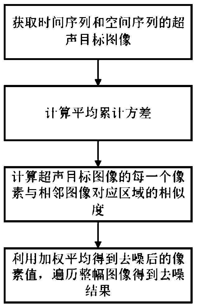
[0038] like figure 1 A method for denoising a human embryonic cardiac ultrasound image is shown, which specifically includes the following steps:

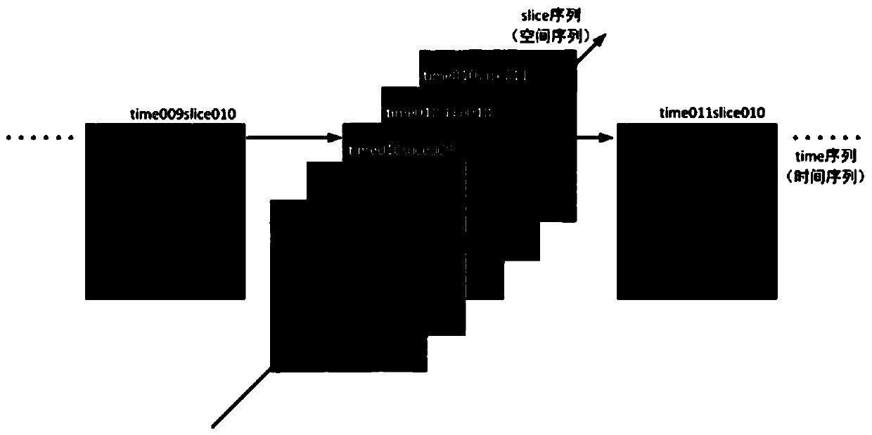
[0039] S1: Acquire an ultrasound image dataset with temporal and spatial sequence features and select a central image to determine the adjacent images of the central image, such as figure 2 shown;

[0040] S11: Convert the ultrasound image contained in the case data into a grayscale image;

[0041] S12: Select an image as the center image, and the original image of the center image is as follows image 3 As shown, two adjacent images in the time series and two adjacent images in the spatial sequence are obtained as adjacent images...


PUM


Abstract

The invention discloses a human embryo cardiac ultrasonic image denoising method which comprises the following steps: S1, acquiring an ultrasonic image data set with time sequence and space sequence characteristics, selecting a central image, and determining an adjacent image of the central image; s2, marking a current pixel to be processed in the central image as a central pixel, calculating thesimilarity between the central pixel and each pixel in the search domain corresponding to the adjacent image; s3, calculating a central pixel gray value corresponding to the adjacent image according to the similarity, and carrying out average value operation on the central pixel gray value to obtain a final gray value of the central pixel; and calculating a corresponding final gray value of each pixel of the central image by adopting the method, and traversing the whole central image to obtain a denoised clear image. According to the method, parameters can be freely adjusted in the denoising process, the denoising effect and time efficiency are balanced, support of a training set is not needed, programming is easy to achieve, and the algorithm complexity is low.

Description

technical field [0001] The invention relates to the technical field of image processing, in particular to a method for denoising a human embryonic cardiac ultrasound image. Background technique [0002] At present, ultrasound technology is an important means to detect whether the fetal heart is healthy, but due to the particularity of the fetus, the ultrasound needs to additionally pass through the fetal mother's abdominal fat to image the embryonic heart, which results in the ultrasound image. More artifacts and noise. In view of this situation, the current solutions are as follows. The first is to use unprocessed ultrasound images for diagnosis based on the experience of medical personnel. This method requires very high technical level of medical personnel. High, it will consume a lot of time and energy, and it is easy to cause misdiagnosis; the second is to use the traditional filtering method to remove noise, although this method has a certain denoising effect, but becau...

Claims


Application Information

Patent Timeline
IPC IPC(8): G06T5/00
CPCG06T2207/10132G06T2207/30048G06T5/70
Inventor 刘斌许钊
Owner DALIAN UNIV OF TECH
Learn More