Separation method of EVs (extracellular vesicles)

A separation method and vesicle technology, applied in the biological field, can solve the problems of expensive, expensive ultra-high-speed centrifuges, and low purity of extracellular vesicles

Pending Publication Date: 2019-09-27
SHANGHAI NINTH PEOPLES HOSPITAL SHANGHAI JIAO TONG UNIV SCHOOL OF MEDICINE
View PDF5 Cites 7 Cited by
  • Summary
  • Abstract
  • Description
  • Claims
  • Application Information

AI Technical Summary

Problems solved by technology

At present, the separation methods of extracellular vesicles mainly include the following: 1. Ultracentrifugation speed (Théry C et al.Curr Protoc Cell Biol, 2006; 3:1-29) or density gradient centrifugation (Tauro BJ et al. Methods 2012; 56:293–304.), this method is considered to be the gold standard for extracellular vesicle isolation, but this method requires an expensive ultra-high-speed centrifuge, so it is not easy to popularize
2. Coagulation and sedimentation method, this method can be divided into two categories, one is to add the extraction reagent to the sample, and centrifuge to obtain the precipitation of extracellular vesicles (LobbRJ et al.J Extracell Vesicles2015; 4:27031), However, the extracellular vesicles extracted by this method are not high in purity and expensive
4. Use cheap polyethylene glycol to precipitate extracellular vesicles (Rider MA et al. Sci Rep 2016; 6:23978). This method is similar to the principle of commercial extracellular vesicle extraction kits. The advantage is that the extraction cost is not high, and the disadvantage is that the purity of the extracted extracellular vesicles is not high

Method used


Image

  • Separation method of EVs (extracellular vesicles)
  • Separation method of EVs (extracellular vesicles)
  • Separation method of EVs (extracellular vesicles)

Examples


Embodiment 1

[0058] 1) Centrifuge 6ml of freshly obtained urine at 10,000rpm for 10min. The precipitate is the cells and impurities in the urine. Remove the precipitate and take 5ml of the supernatant.

[0059] 2) Take 5 mL of the supernatant obtained in step 1) and add it into a 300 kDa dialysis bag. The electrophoresis solution is an aqueous solution of glycine and Tris, and the electrophoresis is performed under the electrophoresis condition of 300mA (Tianneng, EPS600) for 2 hours, and the electrophoresis solution is replaced every 0.5 hours and the electrophoresis direction is changed.

[0060] 3) Add 5ml of the urine extracellular vesicle fluid obtained by dialysis in step 2) (i.e. the solution in the dialysis bag) into a 100kDa ultrafiltration tube, and perform ultrafiltration at 3500rpm for 10min to further concentrate the sample to provide concentrated urine Liquid extracellular vesicles.

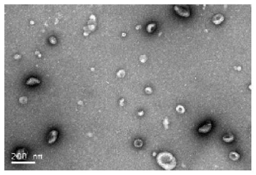
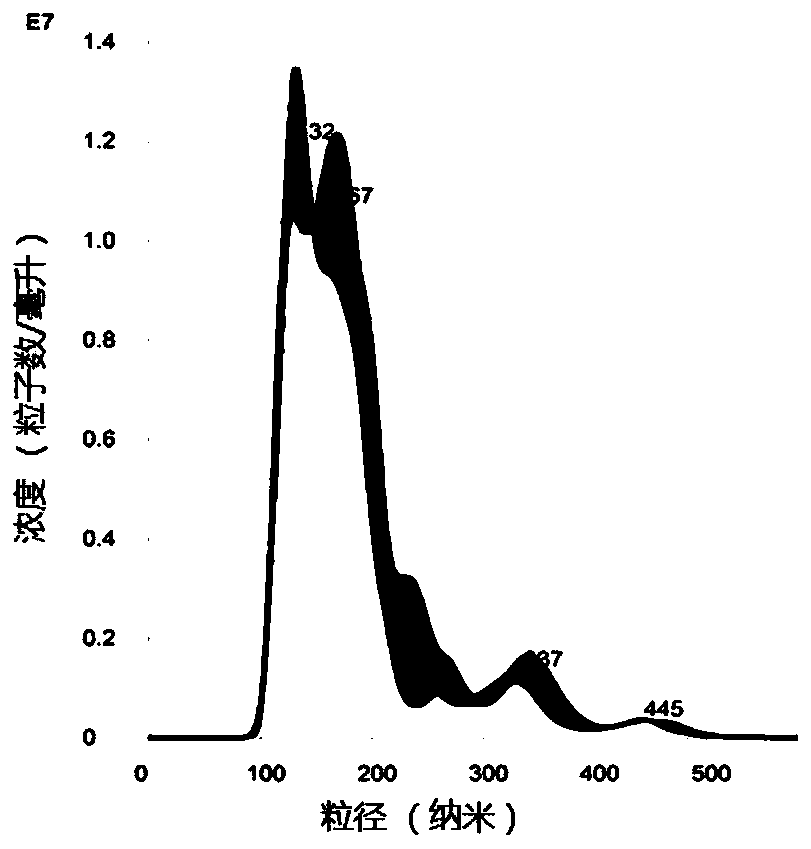
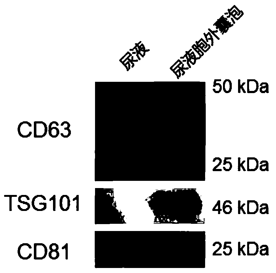
[0061] The obtained urine extracellular vesicles were characterized using a transmission el...

Embodiment 2

[0065] 1) Centrifuge 6ml of freshly obtained saliva at 10,000rpm for 10min. The precipitate is the cells and impurities in the saliva. Remove the precipitate and take 5ml of the supernatant.

[0066] 2) Take 5 mL of the supernatant obtained in step 1) and add it into a 300 kDa dialysis bag. The electrophoresis solution was an aqueous solution of glycine and Tris, and the electrophoresis was performed under the electrophoresis condition of 300mA (Tianneng, EPS 600) for 2 hours, and the electrophoresis solution was changed every 0.5 hours and the electrophoresis direction was changed.

[0067] 3) Add 5ml of the saliva extracellular vesicle fluid obtained by dialysis in step 2) (i.e. the solution in the dialysis bag) into a 100kDa ultrafiltration tube, and perform ultrafiltration at 3500rpm for 10min to further concentrate the sample to provide concentrated saliva Extracellular vesicles.

[0068] The obtained salivary extracellular vesicles were characterized by transmission ele...

Embodiment 3

[0072] 1) Centrifuge the freshly obtained blood at 4000rpm for 10min to obtain serum, and then centrifuge at 10000rpm for 15min to remove the precipitate to obtain the serum supernatant.

[0073] 2) Dilute 0.4 ml of serum supernatant obtained in step 1) to 5 ml with electrophoresis solution.

[0074] 3) Take 5 mL of the diluent obtained in step 2) and add it into a 300 kDa dialysis bag. The electrophoresis solution is an aqueous solution of glycine and Tris, and the electrophoresis is performed for 2.5 hours under the electrophoresis condition of 300mA (Tianneng, EPS600), and the electrophoresis solution is replaced every 0.5 hours and the electrophoresis direction is changed.

[0075] 4) Add 5ml of the serum extracellular vesicle fluid (ie the solution in the dialysis bag) obtained by dialysis in step 3) into a 100kDa ultrafiltration tube, and perform ultrafiltration at 3500rpm for 10min to further concentrate the sample to provide concentrated serum Extracellular vesicles. ...


PUM

PropertyMeasurementUnit
particle diameteraaaaaaaaaa

Abstract

The invention relates to the technical field of biology, in particular to a separation method of EVs (extracellular vesicles). The separation method of the EVs comprises steps as follows: 1) EV samples are subjected to centrifugal treatment to provide a liquid supernatant; 2) the liquid supernatant provided in the step 1) is subjected to electrophoresis dialysis treatment to provide an EV liquid; 3) the EV liquid provided in the step 2) is subjected to ultrafiltration treatment. The EVs are prepared and obtained with the provided separation method, the complete structure of the EVs can be kept, the prepared EVs have the advantages of high purity, low impure protein pollution and the like while multiple samples can be treated simultaneously, almost all EVs in to-be-treated samples can be obtained, and the method has good yield. Besides, the provided method comprises simple steps and is easy to operate, no expensive equipment is involved in the whole process, used consumable materials can be repeatedly recycled, and the method is a low-cost and efficient treatment method.

Description

technical field [0001] The invention relates to the field of biotechnology, in particular to a method for isolating extracellular vesicles. Background technique [0002] Extracellular vesicles (exosomes) are membranous vesicles that can be produced and secreted by almost all living cells, and have important applications in the diagnosis and treatment of diseases. [0003] The isolation of extracellular vesicles is critical for their downstream applications. At present, the separation methods of extracellular vesicles mainly include the following: 1. Ultracentrifugation speed (Théry C et al.Curr Protoc Cell Biol, 2006; 3:1-29) or density gradient centrifugation (Tauro BJ et al. Methods 2012; 56:293–304.), this method is considered to be the gold standard for extracellular vesicle isolation, but this method requires an expensive ultra-high-speed centrifuge, so it is not easy to popularize. 2. Coagulation and sedimentation method, this method can be divided into two categorie...

Claims


Application Information

Patent Timeline
Patent Type & Authority Applications(China)
IPC IPC(8): C12N5/07C12N5/04
CPCC12N5/0601C12N5/0602C12N5/04C12N2509/00
Inventor 杨蒙陈福祥
Owner SHANGHAI NINTH PEOPLES HOSPITAL SHANGHAI JIAO TONG UNIV SCHOOL OF MEDICINE
Why Eureka
Try Eureka