Molecular Imaging Biomarkers

a biomarker and molecular imaging technology, applied in the field of molecular imaging biomarkers, can solve the problems of critical limitation, critical limitation, and non-linearity of stain-free non-linear imaging techniques, and achieve enhanced local invasion, increased photodamage risk, and promotion of tumor malignancy

Active Publication Date: 2018-10-04
THE BOARD OF TRUSTEES OF THE UNIV OF ILLINOIS
View PDF0 Cites 7 Cited by
  • Summary
  • Abstract
  • Description
  • Claims
  • Application Information

AI Technical Summary

Benefits of technology

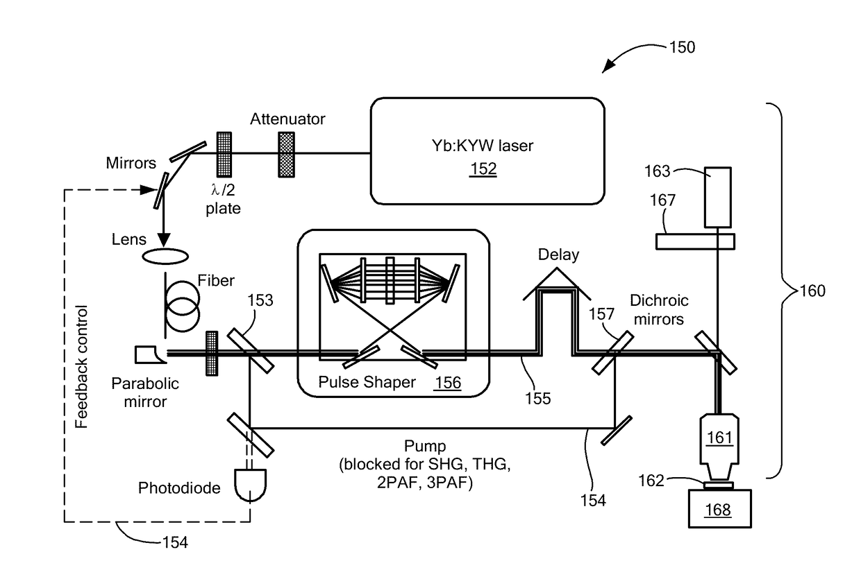
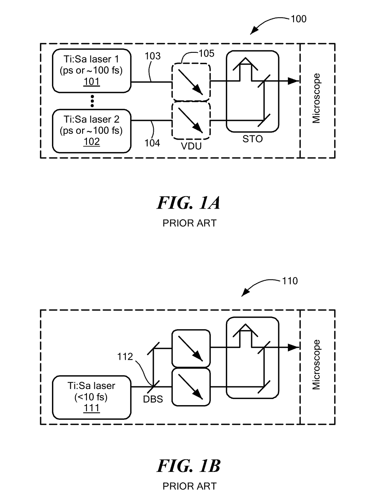
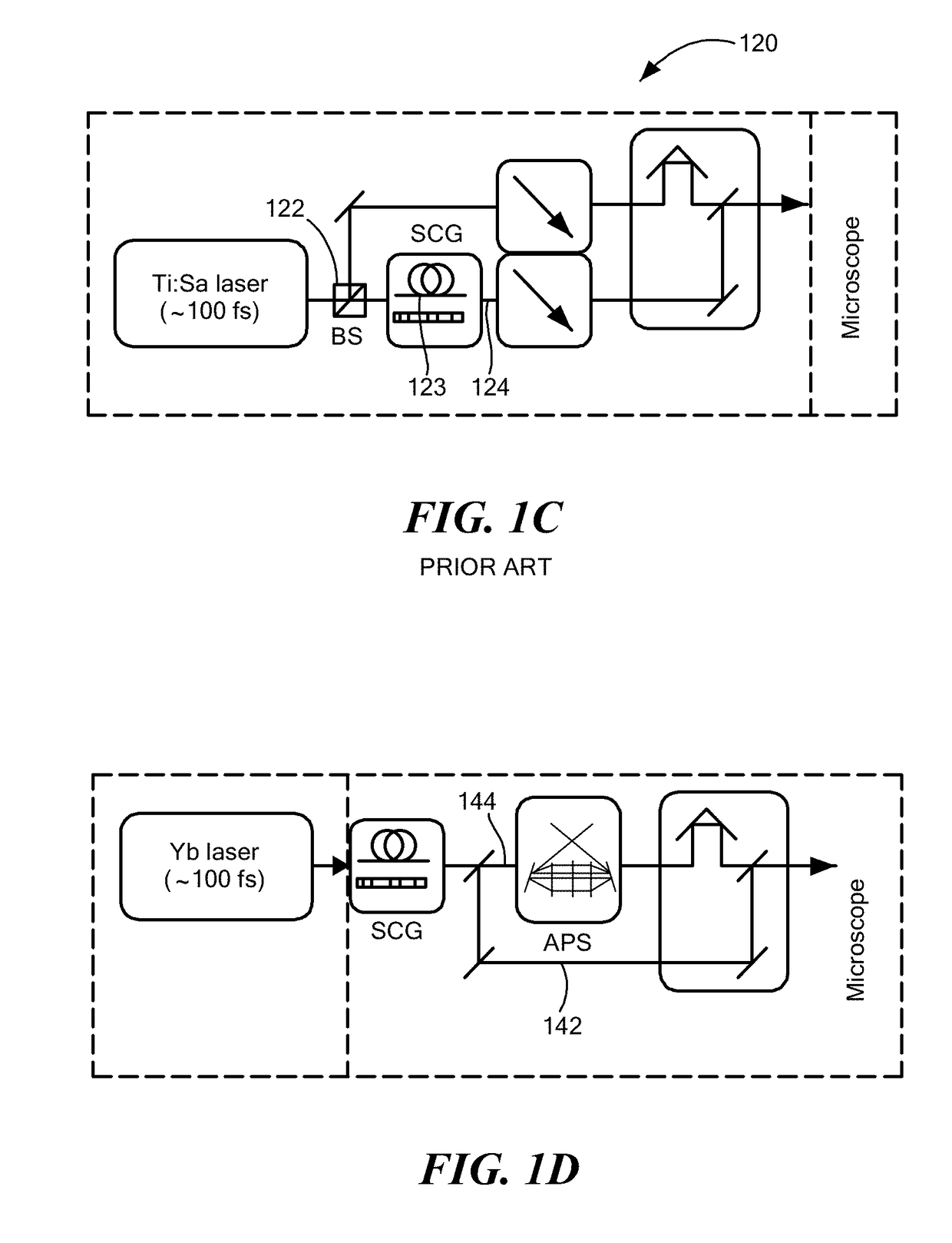
[0011]The former platform has been critically limited by highly nonlinear photodamage, and, to a lesser degree, insufficient structural information when tissue lacks noncentrosymmetry (SHG contrast), while the later platform has been critically limited by the absence of functional information including auto-fluorescence intensity and lifetime. The two platforms complement each other, but have long resisted a synergistic integration to simultaneously collect SHG, THG, and auto-fluorescence signals by one single (single-beam fixed-wavelength) excitation. Their simple combination using sequential SW and LW excitations coupled with sequential signal detections on the same field-of-view, as practiced, for example, in Weigelin et al., “Intravital third harmonic generation microscopy of collective melanoma cell invasion: principles of interface guidance and microvesicle dynamics,” Intravital, vol. 1, pp. 32-43 (2012), incorporated herein by reference, not only increases photodamage risk and prevents rigorous spatial co-registration between sequentially detected signals, but also complicates the corresponding fiber-based endoscope or handheld probe afforded by both platforms. Because THG contrast cannot be generated from the SW-SHG&2PAF platform due to UV absorption by tissue and standard optics, there has long been a profound need to introduce functional information to the LW-SHG&THG platform in order to empower multiphoton microscopy as real-time stain-free histology.
[0012]Various treatments of the tumor microenvironment in the prior literature have included a non-reductionist view of cancer, interactions at the tumor-host interface, a wound-healing analogue of tumor development, the concept of “seed and soil”, the bipolar effects of stroma in the tumor “organ”, and the Darwinian (environmental) selection of metastatic tumor cells. Signature events occurring in the tumor microenvironment include: (I) recruitment or infiltration of non-native cells such as immune inflammatory cells and bone-marrow-derived cells, and activation or alteration of fibroblasts (or other native cells for promotion of tumor malignancy and protection from immune attack; (II) a mechanically reorganized extracellular matrix, including degraded basement membrane and rearranged, cross-linked, or fibrotic collagen for enhanced local invasion; (III) angiogenesis and lymphangiogenesis for primary tumor growth and subsequent metastasis; (IV) modulation of stroma by small (<1 μm) tumor-associated extracellular vesicles for pre-conditioning proliferation, invasion, and metastasis of tumor cells; and (V) metabolic switch from energy production to biomass production (biosynthesis), i.e., a reinterpreted Warburg effect that enriches amino acids, nucleotides, and fatty acids, discussed by Vander Heiden et al., “Understanding the Warburg effect: the metabolic requirements of cell proliferation,” Science, vol. 324, pp. 1029-33 (2009), which is incorporated herein by reference.

Problems solved by technology

The stain-free non-linear imaging techniques obviate time-consuming paradigmatic elements of standard histology procedures.
The former platform has been critically limited by highly nonlinear photodamage, and, to a lesser degree, insufficient structural information when tissue lacks noncentrosymmetry (SHG contrast), while the later platform has been critically limited by the absence of functional information including auto-fluorescence intensity and lifetime.
32-43 (2012), incorporated herein by reference, not only increases photodamage risk and prevents rigorous spatial co-registration between sequentially detected signals, but also complicates the corresponding fiber-based endoscope or handheld probe afforded by both platforms.
However, the interrelation between the events at the macroscopic scale (I-III) and microscopic scale (IV, V) remains elusive due to the lack of an imaging methodology to observe them in concert, in spatially-resolved ways, and without perturbative labels.
However, the molecular specificity, which is crucial in the era of molecular oncology, has not been satisfactory using the linear microscopy techniques.

Method used


Image

  • Molecular Imaging Biomarkers
  • Molecular Imaging Biomarkers
  • Molecular Imaging Biomarkers

Examples


example i

n-Induced Rat Mammary Tumor Model

[0097]A well-known preclinical carcinogen-induced rat mammary tumor model was used, and a mammary tumor / tissue specimen (5×5 mm2 area with ˜2 mm thickness) was excised from a rat six weeks after carcinogen injection. The collection of the multimodal multiphoton images began within minutes after dissection from a site with cleanly delineated adipose and stromal regions, and standard H&E histology was subsequently performed to locate an anatomically similar site for comparison. FIGS. 2A-2C illustrate the mesoscopic organization of biological microstructures revealed in co-localized multiphoton images of two rat mammary specimens and absent from corresponding FFPE-H&E histology images, as now described. Area-integrated CARS spectra over 34 hyperspectral images confirm the presence of a significant R3050 peak as a potential cancer biomarker. FIG. 2A shows that different histochemical components of a tumor are selectively revealed by different single-moda...

example ii

ve Breast Cancer Biomarkers

[0101]A potential quantitative breast cancer biomarker was demonstrated for the first time in the rat model discussed above. It was discovered that tumors can be objectively discriminated against non-tumor specimens by the emergence of an R3050 spectral peak in the hyperspectral image-integrated CARS spectra, shown in FIGS. 2A-2C and 8. This R3050 signal is not from the carcinogen (N-nitroso-N-methylurea) injected into the rats because spectral focusing of CARS on a saturated water solution of the carcinogen (1.4% by weight) did not yield any signal at R3050. Because regions within mammary specimens can be largely classified into stromal regions (FIG. 7 and adipose regions (FIG. 9) that account for 25% and 75% of the total area from control specimens, respectively, it is more representative to examine comparable tumor and non-tumor specimens that contain both regions.

[0102]FIG. 9 combines co-localized single-modality multiphoton images in mammary adipose r...

example iii

of Extracellular Vesicle Enrichment and Metabolic Switch Visualized Label-Free in the Tumor Microenvironment

[0112]The in vivo role of extracellular vesicles has been relatively elusive without observing them label-free in tissue. By visualizing these vesicles in situ in the unperturbed tumor microenvironment, the present inventors have found a direct link of their enrichment with the metabolic switch toward biosynthesis. Thus, these vesicles may serve as the signaling mediators from the tumor cells to the abundant stromal cells in order to initiate the metabolic switch, which may in turn induce various macroscopic events in the tumor microenvironment. Genetic modification of the stromal cells can occur when they accept the vesicles with RNA, according to J. Skog et al., “Glioblastoma microvesicles transport RNA and proteins that promote tumour growth and provide diagnostic biomarkers,” Nat. Cell Biol., vol. 10, pp. 1470-76 (2008), incorporated herein by reference. The observations m...


PUM

PropertyMeasurementUnit
diameteraaaaaaaaaa
coupling poweraaaaaaaaaa

Abstract

Methods for label-free characterization of untagged molecules within a biological sample in-situ. The untagged molecules may be constituent of extracellular vesicles, and are excited in the biological sample with at least one wavelength band of light derived from a single stream of optical pulses. Light emitted by the untagged molecules by SHG, THG, 2PAF and 3PAF processes is detected. Separate measures of the biological sample corresponding to light emitted by the untagged molecules in each of the SHG, THG, 2PAF and 3PAF processes are derived. On that basis, normal extracellular vesicles may be differentiated from extracellular vesicles associated with a tumor on the basis of a specified signature of characteristics of images of SHG, THG, 2PAF and 3PAF processes.

Description

[0001]The present application claims priority from U.S. Provisional Patent Application Ser. No. 62 / 478,355, filed Mar. 29, 2017, and incorporated herein by reference.[0002]This invention was made with government support under grant CA166309 awarded by the National Institutes of Health. The government has certain rights in the invention.TECHNICAL FIELD[0003]The present invention relates to apparatus and methods for in-situ label-free imaging of molecules, and, more particularly, molecules associated with extracellular vesicles using concurrent multimodal nonlinear imaging.BACKGROUND ART[0004]Over the course of recent years, stain-free histopathology of fresh tissue within minutes has been established by diverse nonlinear optical processes to improve upon conventional histotechnology based on single-photon microscopy. The stain-free non-linear imaging techniques obviate time-consuming paradigmatic elements of standard histology procedures. A summary of various of the nonlinear optical...

Claims


Application Information

Patent Timeline
Patent Type & Authority Applications(United States)
IPC IPC(8): G06T7/00G01N33/483G06F19/24G01N21/65G01N21/64G16B40/10
CPCG06T7/0014G01N33/483G06F19/24G01N21/65G01N21/6486G01N2201/06113G06T2207/30024G06T2207/30096G06T2207/10064G06T2207/20081G01N21/636G01N2021/653G01N15/1456G01N2015/144G01N2015/1006G16B40/00G16B40/10
Inventor BOPPART, STEPHEN A.TU, HAOHUAYOU, SIXIANLIU, YUAN
Owner THE BOARD OF TRUSTEES OF THE UNIV OF ILLINOIS
Why Eureka
Try Eureka