

Immunohistochemical staining kit containing pigment tissues and staining method
A technology of immunohistochemistry and staining reagents, applied in the field of immunostaining, which can solve the problems of affecting the interpretation of immunohistochemistry results, covering the morphology and structure of tissue cells, and low sensitivity of the detection system, so as to achieve clear and distinguishable immunohistochemistry results and staining background. Clean, signal-clear results
- Summary
- Abstract
- Description
- Claims
- Application Information
AI Technical Summary
Problems solved by technology
Method used
Image

Examples
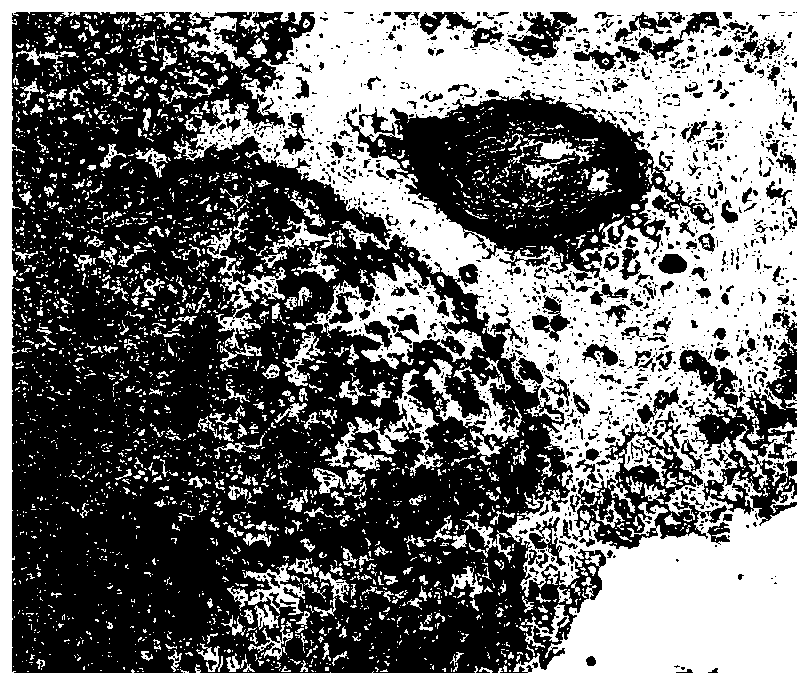
Embodiment 1
[0049] In this embodiment, the method for immunohistochemical staining of pigmented tissue comprises the following steps:
[0050] 1) Take the pigmented tissue section to be tested and add peroxidase blocking agent dropwise to block endogenous peroxidase;
[0051] 2) Incubate the tissue sections with the target antibody, and then incubate with the peroxidase-labeled secondary antibody polymer;
[0052] 3) Incubate and stain the tissue sections at room temperature for 5-10 minutes using the blue dye chromogenic solution; the blue dye chromogenic solution includes: the blue dye substrate based on the peroxidase system and the blue dye that matches the blue dye substrate buffer;
[0053] 4) Use the red contrast agent to stain the tissue sections.
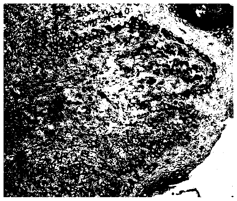
Embodiment 2
[0055] In this embodiment, the method for immunohistochemical staining of pigmented tissue comprises the following steps:
[0056] 1) Take one melanoma tissue, prepare 6 paraffin tissue sections with a thickness of 3 microns, and bake the slices at 60°C for 1 hour.
[0057] 2) Routine dewaxing and hydration of tissue sections: incubate xylene Ⅰ and Ⅱ for 15 minutes each, ethanol Ⅰ and Ⅱ for 5 minutes each, 95% ethanol, 80% ethanol and 70% ethanol for 5 minutes each, Wash with pure water, soak for 3 minutes*3 times.
[0058] 3) Antigen restoration: use Tris-EDTA (pH9.0) restoration solution, repair under high pressure for 3 minutes, cool naturally, wash with PBST, soak for 3 minutes*3 times.
[0059] 4) Blocking with blocking agent: Incubate with peroxidase blocking agent to block endogenous peroxidase; 100 microliters of reagent per slice, incubate at room temperature for 5 minutes, wash with PBST buffer, soak for 3 minutes*3 times.
[0060] 5) Antibody incubation: Sections ...
Embodiment 3
[0068] In this embodiment, the method for immunohistochemical staining of pigmented tissue comprises the following steps:
[0069] 1) Take one melanoma tissue and one lung adenocarcinoma tissue, prepare two paraffin tissue sections with a thickness of 3 microns, and bake the slices at 60°C for 1 hour.
[0070] 2) Routine dewaxing and hydration of tissue sections: incubate xylene Ⅰ and Ⅱ for 15 minutes each, ethanol Ⅰ and Ⅱ for 5 minutes each, 95% ethanol, 80% ethanol and 70% ethanol for 5 minutes each, Wash with pure water, soak for 3 minutes*3 times.
[0071] 3) Antigen restoration: use Tris-EDTA (pH9.0) restoration solution, repair under high pressure for 3 minutes, cool naturally, wash with PBST, soak for 3 minutes*3 times.
[0072] 4) Blocking with blocking agent: Incubate with peroxidase blocking agent to block endogenous peroxidase; 100 microliters of reagent per slice, incubate at room temperature for 5 minutes, wash with PBST buffer, soak for 3 minutes*3 times.
[00...
PUM
 Abstract
Description
Claims
Application Information