Reagent kit for detecting tuberculous pleurisy

A tuberculous pleurisy and kit technology, applied in the field of biomedicine, can solve the problems of difficult diagnosis, tuberculosis dissemination, and low sensitivity

Inactive Publication Date: 2019-10-18
BEIJING TUBERCULOSIS & THORACIC TUMOR RES INST +1
View PDF1 Cites 0 Cited by
  • Summary
  • Abstract
  • Description
  • Claims
  • Application Information

AI Technical Summary

Problems solved by technology

Bacteriology is the gold standard for the diagnosis of tuberculous pleurisy, but the diagnosis is very difficult
Tuberculous pleurisy is extrapulmonary tuberculosis, the diseased site is in the middle of the pleura, bacteria are often not expelled from the body through sputum, and the sensitivity of finding acid-fast bacilli is extremely low (0% to 5%); a large amount of pleural effusion in patients with tuberculous pleurisy The concentration of Mycobacterium tuberculosis was reduced, resulting in only 5.8% positive rate of pleural fluid smear, although the positive rate of modified Roche culture was increased (30%), but the culture time was longer (4-8 weeks); the positive rate of pleural biopsy The rate reaches 69.6-82%, but the trauma is relatively large, and there is a risk of causing the spread of tuberculosis and exacerbating the disease

Method used


Image

  • Reagent kit for detecting tuberculous pleurisy
  • Reagent kit for detecting tuberculous pleurisy
  • Reagent kit for detecting tuberculous pleurisy

Examples


Embodiment 1

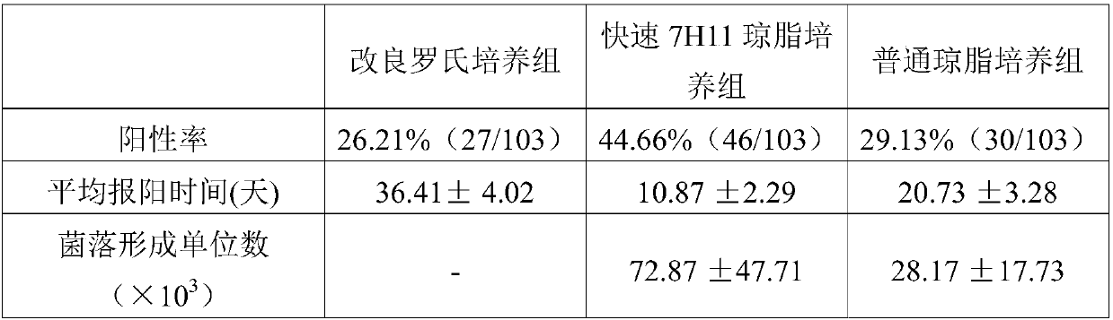
[0048] Example 1. Detection of Mycobacterium tuberculosis in pleural effusion

[0049] 1. Experimental method

[0050] 1. Specimen processing

[0051] Aseptically extract 50-200m1 of pleural effusion from each patient with tuberculous pleurisy (a total of 103 samples, all of whom were clinically diagnosed with tuberculous pleurisy, and the patients were informed and agreed to this experiment), and packed in 50m1 sterile centrifuge tubes. After centrifuging at 3000×g for 20 minutes, discard part of the supernatant, leave 5ml at the bottom of the tube, add an equal volume of 4g / 100ml sodium hydroxide (NaOH), vortex, wait for the sample to be uniformly liquefied and let stand at 20-25°C for 15 minutes. Add 30ml of 0.067mol pH 6.8 phosphate buffer and mix well; centrifuge at 3000g for 30min, discard the supernatant, add 0.5m1 0.067mol pH 6.8 phosphate buffer and mix well to obtain the processed specimen, and set aside.

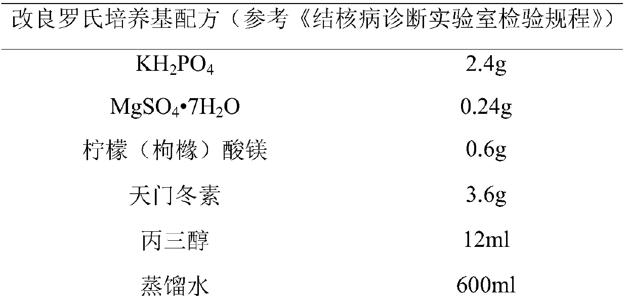
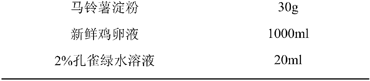
[0052] 2. Preparation of 7H11 agar medium

[0053] Add 2....

Embodiment 2

[0095] Example 2, the results of different Mycobacterium tuberculosis recovery factor proteins in the rapid 7H11 agar culture group

[0096] The present invention explores the mixed molar ratio concentrations of recovery factors RpfA protein, RpfB protein, RpfC protein, RpfD protein and RpfE protein (see Table 3). The experimental method is carried out with reference to Example 1. The results showed that the effect of scheme ②-⑦ was significantly better than that of scheme ① in terms of positive rate, average positive reporting time (days) and number of colony forming units.

[0097] Table 3 Comparison of positive results of pleural effusion in 7H11 agar culture groups with different molar ratio concentrations of recovery factors

[0098]

[0099] Note: ①Scheme (Rpf B: 137nmol, Rpf E: 121nmo1);

[0100] ② Scheme (Rpf B: 13.7nmol, Rpf C: 47nmo1);

[0101] ③ Scheme (Rpf A: 15.6nmol, Rpf E: 12.1nmo1);

[0102] ④ Scheme (Rpf A: 156nmol, Rpf B: 1.4nmol, Rpf C: 47nmo1, Rpf D:...


PUM

PropertyMeasurementUnit
thicknessaaaaaaaaaa

Abstract

The invention discloses a reagent kit for detecting tuberculous pleurisy, provides a reagent kit for detecting whether mycobacterium tuberculosis exists in hydrothorax, and relates to a recovery factor protein composition containing a 7H11 solid culture medium and the mycobacterium tuberculosis. The recovery factor protein composition consists of at least two of RpfA protein, RpfB protein, RpfC protein, RpfD protein and RpfE protein, wherein the mol parts of the proteins conforms to the following standard: 0-156 parts of the RpfA protein, 0-173 parts of the RpfB protein, 0-473 parts of the RpfC protein, 0-150 parts of the RpfD protein, and 0-121 parts of the RpfE protein. Experiment proves that the reagent kit for detecting mycobacterium tuberculosis in hydrothorax has the characteristicsof being short in positive report time, high in positive rate, and high in formation unit number of positive colonies. The reagent kit has important significance in detection or auxiliary detection ofthe tuberculous pleurisy.

Description

technical field [0001] The invention belongs to the field of biomedicine and relates to a kit for detecting tuberculous pleurisy. Background technique [0002] my country is a country with a high burden of tuberculosis, and tuberculous pleurisy is a common cause of pleural effusions, accounting for about 50% of pleural effusions. Bacteriology is the gold standard for the diagnosis of tuberculous pleurisy, but the diagnosis is very difficult. Tuberculous pleurisy is extrapulmonary tuberculosis, the diseased site is in the middle of the pleura, bacteria are often not expelled from the body through sputum, and the sensitivity of finding acid-fast bacilli is extremely low (0% to 5%); a large amount of pleural effusion in patients with tuberculous pleurisy The concentration of Mycobacterium tuberculosis was reduced, resulting in only 5.8% positive rate of pleural fluid smear, although the positive rate of modified Roche culture was increased (30%), but the culture time was longe...

Claims


Application Information

Patent Timeline
Patent Type & Authority Applications(China)
IPC IPC(8): C12Q1/04C12N1/20C12N1/02C12R1/32
CPCC12Q1/04C12N1/20C12N1/02
Inventor 刘忠泉邢爱英杜凤娇张宗德
Owner BEIJING TUBERCULOSIS & THORACIC TUMOR RES INST
Why Eureka
Try Eureka