
Method for quantitative detection of single embryo secreted proteins based on microfluidic droplets

A quantitative detection method and technology of secreted protein, which is applied in the field of quantitative detection of single embryo secreted protein based on microfluidic droplets, can solve the problem of inability to accurately detect secreted protein of a single embryo, and reduce the interference of human subjective factors and achieve high detection efficiency. Sensitivity, effect of reducing reaction time

Active Publication Date: 2019-10-22
深圳中山泌尿外科医院
View PDF3 Cites 4 Cited by
  • Summary
  • Abstract
  • Description
  • Claims
  • Application Information

AI Technical Summary

Problems solved by technology

[0005] In order to solve the technical problem of being unable to accurately detect the secreted protein of a single embryo in the above background technology, the present invention provides a quantitative detection method for the secreted protein of a single embryo based on microfluidic liquid droplets, which can accurately detect the secreted protein of a single embryo

Method used


Image

  • Method for quantitative detection of single embryo secreted proteins based on microfluidic droplets
  • Method for quantitative detection of single embryo secreted proteins based on microfluidic droplets
  • Method for quantitative detection of single embryo secreted proteins based on microfluidic droplets

Examples


Embodiment 1

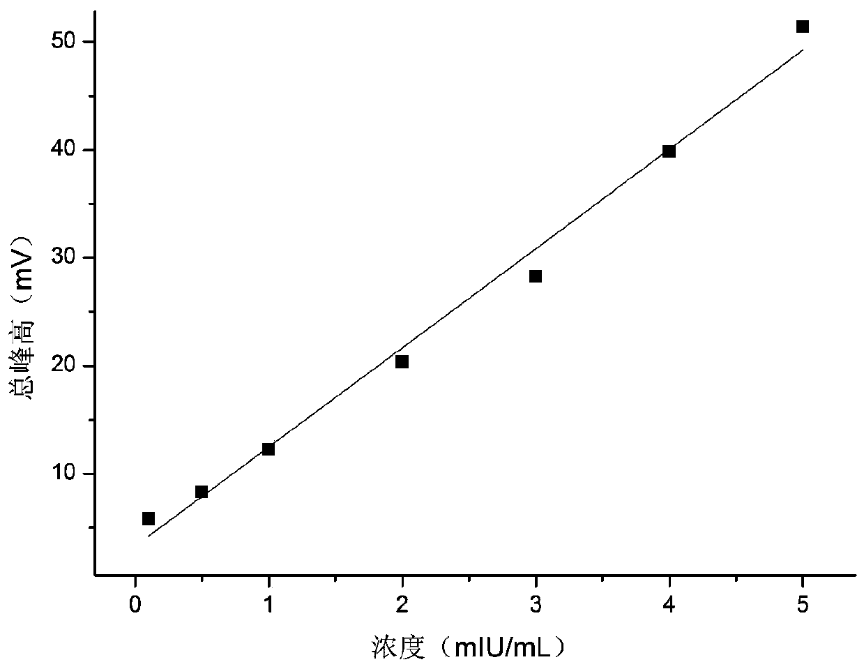
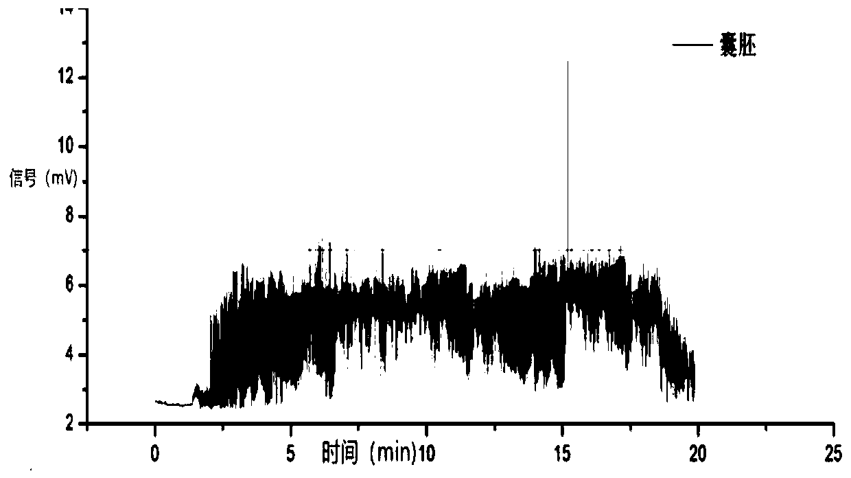
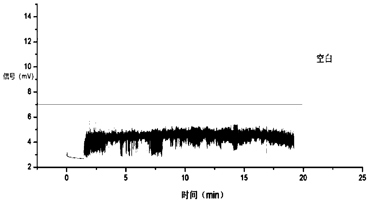
[0030] like figure 1 As shown, a method for quantitative detection of protein secreted by a single embryo based on microfluidic droplets, is characterized in that it comprises the following steps:

[0031] a. Take a capture antibody and a labeled antibody that secrete human chorionic gonadotropin (βHCG) from a single blastocyst stage embryo, connect the capture antibody to immunomagnetic beads, and label the labeled antibody with biotin;

[0032] b. Mix the immunomagnetic beads in step a with human chorionic gonadotropin (βHCG) secreted by a single blastocyst embryo and incubate at 37°C for 30 minutes, then take out the immunomagnetic beads and wash them 3 to 5 times. Mix the magnetic beads with the labeled antibody and streptavidin-labeled β-galactosidase and incubate at 37°C for 30 minutes, then take out the immunomagnetic beads and wash them 3 to 5 times, and resuspend them with 10uL of PBS buffer after washing Immunomagnetic beads;

[0033] c. Put the respun magnetic bea...


PUM

PropertyMeasurementUnit
Diameteraaaaaaaaaa
Excitation wavelengthaaaaaaaaaa

Abstract

The invention, which belongs to the field of detection methods, in particular relates to a method for quantitative detection of single embryo secreted proteins based on microfluidic droplets. The detection method comprises the steps: taking a capture antibody and a label antibody of a to-be-detected single embryo secreted protein, connecting the capture antibody to an immunomagnetic bead and labeling the label antibody by biotin; mixing the immunomagnetic bead with the to-be-detected single embryo secreted protein and carrying out incubation, carrying out cleaning, mixing the mixture with thelabel antibody and streptavidin-labeled beta-galactosidase and carrying out incubation, and re-suspending the immunomagnetic bead by using a PBS buffer solution; and then detectint he fluorescence intensity by a laser-induced fluorescence detector to obtain the content of the to-be-detected single embryo secreted protein. According to the invention, substrate reaction is catalyzed continuously byusing an enzymatic reaction to generate a fluorescent substance, so that the signal amplification of the to-be-detected protein is realized; and with introduction of the microfluidic droplet technology, the fluorescent substances are enriched quickly to reach the measurable concentration. The detection sensitivity is improved; and the size of the sampled needed by detection is reduced.

Description

technical field [0001] The invention belongs to the field of detection methods, and in particular relates to a method for quantitative detection of single embryo secreted protein based on microfluidic liquid droplets. Background technique [0002] A variety of proteins are secreted during embryonic growth and development, and these proteins are closely related to various functions during embryonic development and implantation. A single embryo refers to an embryo, and the detection of the secreted protein content of a single embryo in a single embryo culture medium is of great significance for understanding the development of the embryo and exploring the developmental potential of the embryo. Single-embryo secreted protein refers to the protein content of secreted protein in a single embryo culture medium. However, the concentration of protein secreted by a single embryo is extremely low, and the volume of culture medium is only 20-30 μL. At the same time, protein cannot be ...

Claims


Application Information

Patent Timeline
IPC IPC(8): G01N21/64G01N33/76G01N33/68
CPCG01N21/6402G01N21/6428G01N21/6486G01N33/76G01N33/689
Inventor 陈培林李观贵孙青熊风钟惠娴姚志鸿
Owner 深圳中山泌尿外科医院
Learn More