Method for detecting content of proximal renal tubule source exosome in urine

A detection method and technology of renal tubules, which are applied in the field of medical detection, can solve the problems of insufficient application value, neglect of the importance of renal tubular lesions, and difficulties in subsequent detection of protein markers, so as to achieve the effect of increasing feasibility and achieving content

Inactive Publication Date: 2019-11-05
THE SECOND AFFILIATED HOSPITAL OF NANJING MEDICAL UNIV
View PDF3 Cites 2 Cited by
  • Summary
  • Abstract
  • Description
  • Claims
  • Application Information

AI Technical Summary

Problems solved by technology

[0005] However, at present, the traditional diagnosis and classification methods of kidney diseases still focus on glomerular lesions, often ignoring the importance of renal tubular lesions for disease progression and prognosis
In addition, although the exosome was discovered earlier, related research has only gradually received attention in recent years, and the theoretical basis and research methods are still being improved. The understanding of the application value of cell-derived exosomes in disease

Method used


Image

  • Method for detecting content of proximal renal tubule source exosome in urine
  • Method for detecting content of proximal renal tubule source exosome in urine
  • Method for detecting content of proximal renal tubule source exosome in urine

Examples


Embodiment 1

[0049] A method for detecting exosome content derived from proximal renal tubules in urine, comprising the following steps:

[0050] S1, preparation of urine exosome suspension

[0051] (1) Collect 100ml of fresh morning urine from healthy subjects, centrifuge at 3000g for 10min, and collect the supernatant A;

[0052] (2) Add 8.44ml protease inhibitor mixture to the supernatant A, mix well and centrifuge at 17000g for 10min at 4°C to collect the supernatant B;

[0053] Wherein, the protease inhibitor mixture is formed by mixing sodium azide, phenylmethylsulfonyl fluoride, and leupeptin in a molar ratio of 100:10:1;

[0054] (3) The supernatant B was centrifuged at 20°C and 200,000g for 1.5h, the precipitate D was collected, and the precipitate D was resuspended with 100 μl of sucrose separation solution to obtain a resuspension;

[0055] Wherein, the sucrose separation solution is formed by mixing triethanolamine and sucrose at a molar ratio of 1:25;

[0056] (4) Add dithi...

Embodiment 2

[0078] A method for detecting exosome content derived from proximal renal tubules in urine, comprising the following steps:

[0079] S1, preparation of urine exosome suspension

[0080] The specific steps are the same as in Example 1, except that 100 ml of fresh morning urine from patients with proximal renal tubular lesions was used to prepare the urine exosome suspension;

[0081] S2, Determination of protein concentration in urine exosome suspension

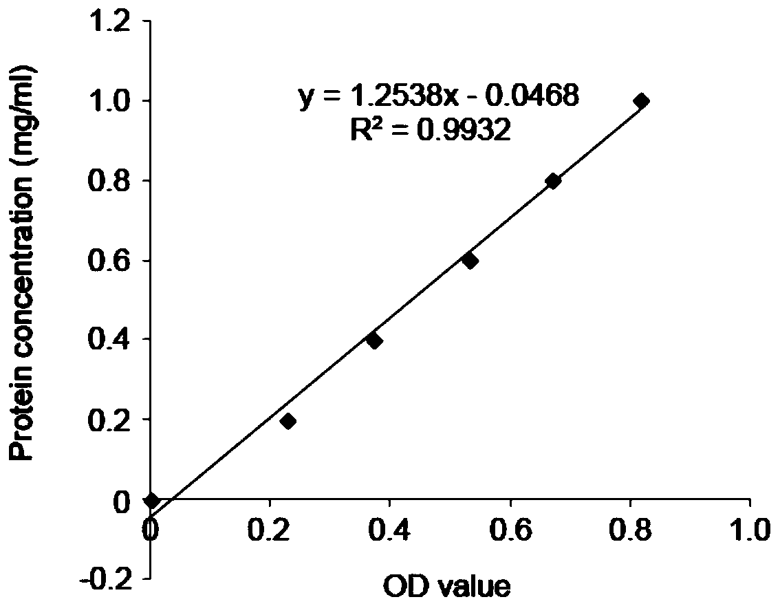
[0082] The specific steps are the same as in Example 1, the difference is that the OD value read during the detection of the protein concentration in the exosome lysate is 0.362, and the OD value is brought into the above linear regression equation to calculate the protein concentration in the exosome lysate, exosome The protein concentration in the lysate is (1.2538×0.362-0.0468)×5 (dilution factor)=2.035mg / ml, which is the protein concentration in the urine exosome suspension;

[0083] S3, the exosome derived from the prox...


PUM


Abstract

The invention discloses a method for detecting the content of a proximal renal tubule source exosome in urine. The method includes the following steps of S1, preparing urine exosome suspension; S2, measuring the protein concentration in the urine exosome suspension; S3, separating out the proximal renal tubule source exosome in the urine exosome suspension; S4, detecting the proximal renal tubulesource exosome separated out of urine in the step S3. The high-purity urine exosome is obtained through an ultracentrifugation method, the exosome is then adsorbed by immunomagnetic beads, the problemis solved that the exosome is small in size, and the feasibility of detecting the exosome through a flow cytometry method is increased; the CD63 and CD13 immunolabelling method is adopted for separating out the proximal renal tubule source exosome in the urine and subsequently accurately detecting and analyzing the content of the proximal renal tubule source exosome in the urine.

Description

technical field [0001] The invention relates to the technical field of medical detection, in particular to a method for detecting the content of exosome derived from proximal renal tubules in urine. Background technique [0002] With the improvement of people's living standards and the development of medical and health services, the prevalence of various chronic diseases has increased year by year, especially hypertension, diabetes and chronic kidney disease. The number of patients in my country has reached hundreds of millions. . Traditional renal disease assessment methods mainly include serology, urine biochemical and imaging examinations, such as serum urea nitrogen and creatinine, routine examinations such as urine protein and occult blood, and urinary system ultrasound, which have poor sensitivity and specificity, such as serum urea Nitrogen and creatinine will only become abnormal when the renal function is lost by more than 50%, and urine protein and occult blood wil...

Claims


Application Information

Patent Timeline
IPC IPC(8): G01N33/68G01N33/543G01N15/10G01N21/78G01N21/31
CPCG01N15/10G01N21/31G01N21/78G01N33/54326G01N33/6893
Inventor 杨俊伟周阳秦楠徐玲玲江蕾曹红娣熊明霞闻萍
Owner THE SECOND AFFILIATED HOSPITAL OF NANJING MEDICAL UNIV
Why Eureka
Try Eureka