Puncture forceps holder biopsy needle used beneath bronchoscope

A bronchoscope and biopsy needle technology, applied in the field of medical devices, can solve problems such as inability to penetrate the tracheal wall, blunt tip of biopsy forceps, and inability to reach the mediastinum for biopsy, achieving efficient diagnosis, less discomfort, and convenient operation

Pending Publication Date: 2019-11-22
夏旸
View PDF0 Cites 7 Cited by
  • Summary
  • Abstract
  • Description
  • Claims
  • Application Information

AI Technical Summary

Problems solved by technology

The volume of tissue samples obtained by biopsy forceps is large, but the tip of the biopsy forceps is blunt, which cannot penetrate the tracheal wall and cannot reach the mediastinum for biopsy

Method used


Image

  • Puncture forceps holder biopsy needle used beneath bronchoscope
  • Puncture forceps holder biopsy needle used beneath bronchoscope
  • Puncture forceps holder biopsy needle used beneath bronchoscope

Examples


Embodiment 1

[0012] Embodiment 1: A kind of puncture forceps biopsy needle used under bronchoscope

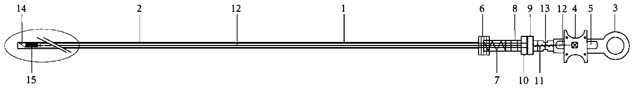
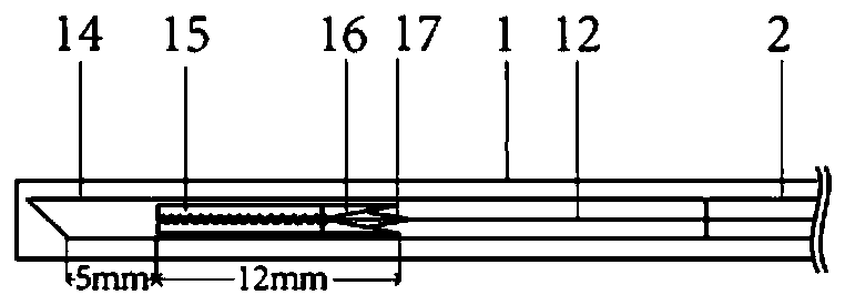
[0013] see figure 1 , figure 2 , a puncture forceps biopsy needle used under a bronchoscope, consisting of an overtube 1, a connecting wire 2, a pull ring 3, a slider 4, a chute 5, a spiral sleeve mouth 6, a spiral interface 7, a sliding handle 8, and a puncture needle Slide block 9, biopsy forceps slide block 10, spring 11, tension steel wire 12, draw-in groove 13, puncture needle 14, biopsy forceps 15, scissors structure 16, dragline 17 form. Among them, one end of the tension steel wire 12 is hinged with the biopsy forceps 15, and the opening and closing of the biopsy forceps is controlled by the change of tension, and one end is hinged with the slider 4, and the position of the biopsy forceps is changed by changing the position of the slider, while the puncture needle slider 9 and the biopsy forceps slide A small hole is set in the center of the block 10 for the tension steel wire 1...

Embodiment 2

[0014] Example 2: Transbronchial Needle Aspiration Biopsy under Common Bronchoscope

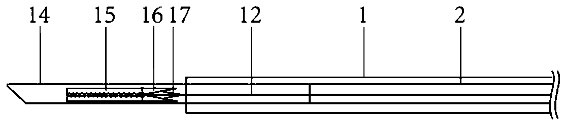
[0015] During routine bronchoscopy, the biopsy needle figure 1 In this state, extend from the working channel of the ordinary bronchoscope into the airway, find the puncture point, turn the helical sleeve 6 at the handle end to coincide with the helical interface 7, thereby retracting the overtube and exposing the puncture needle, such as image 3 , the puncture needle penetrates the bronchial wall and enters the designated lesion. At this time, there is a 5mm gap between the biopsy forceps and the puncture needle tip to accommodate the tissue, and a transbronchial needle aspiration biopsy is possible. After the needle biopsy is completed, the slider 9 is moved to the slot 13, the spring is compressed, the puncture needle is retracted, and the biopsy forceps are exposed. The position structure at this time is as follows: Figure 4 At this time, the biopsy forceps are equivalent to the inner ...

Embodiment 3

[0016] Example 3: Transbronchial clip biopsy under common bronchoscope

[0017] During routine bronchoscopy, the biopsy needle figure 1 In this state, extend from the working channel of the ordinary bronchoscope into the airway, find the puncture point, turn the helical sleeve 6 at the handle end to coincide with the helical interface 7, thereby retracting the overtube and exposing the puncture needle, such as image 3 , the puncture needle penetrates the bronchial wall and enters the designated lesion, move the slider 9 to the slot 13, compress the spring, retract the puncture needle, and expose the biopsy forceps. At this time, the position structure is as follows Figure 4 . Figure 4 In this state, the operator can operate the slide block 4 to move backward, and the drag cable 17 hinged with the tension steel wire 12 will be tense thereupon, which will affect the biopsy forceps to open, and then push the slide block 4 forward to close the mouth of the biopsy forceps cup, ...


PUM


Abstract

The invention provides a puncture forceps holder biopsy needle used beneath a bronchoscope. The puncture forceps holder biopsy needle is composed of an outer sleeve tube, a puncture needle, biopsy forceps, a tensioning steel wire, a connecting steel wire, and a sliding block, a helical casing and a helical connector which correspondingly control the puncture needle and the biopsy forceps, a sliding block and a handle which control the tensioning steel wire and the like. In the outer sleeve tube of the puncture forceps holder biopsy needle, the puncture needle is connected with a puncture needle sliding block through the connecting steel wire, the biopsy forceps are located in the puncture needle and connected with the biopsy forceps sliding block through the tensioning steel wire. According to the puncture forceps holder biopsy needle, transbronchial needle aspiration and forcep biopsy can be achieved on the same puncture part, the quantity of obtained samples can be increased, the diagnostic rate of mediastinal lesions is increased, meanwhile the discomfort of a patient is greatly reduced, and the safety of the patient is improved.

Description

technical field [0001] The invention belongs to the field of medical instruments, in particular to a puncture forceps biopsy needle used under a bronchoscope. Background technique [0002] At present, with the development of bronchoscopy technology, transbronchial needle aspiration (TBNA) and biopsy forceps biopsy have become important means of obtaining tissue samples under bronchoscopy for definite diagnosis. TBNA needles can penetrate the tracheal wall and are mostly used for puncturing mediastinal enlarged lymph nodes, while the latter is mostly used for direct clamping of endotracheal lesion tissue under the microscope. However, the sample size obtained by needle biopsy is small, especially for benign lesions with tough tissues, usually only cytology results are available, which is difficult to meet the current development trend of pathological diagnosis based on multiple immunohistochemistry. In order to obtain tissue, puncture can only be performed repeatedly, which ...

Claims


Application Information

Patent Timeline
Patent Type & Authority Applications(China)
IPC IPC(8): A61B10/04A61B10/06A61B17/34
CPCA61B10/0283A61B10/04A61B10/06A61B17/3403A61B2010/045A61B2017/3413
Inventor 夏旸夏乐欣陈婷李雯沈华浩
Owner 夏旸
Why Eureka
Try Eureka