Integration of sample separation with rapid diagnostic tests for improved sensitivity

a sample separation and rapid diagnostic technology, applied in the field of malaria rdts, can solve the problems of increasing the number of steps required for the assay, affecting the detection performance (sensitivity) of the test kit, false negative test, etc., and achieves the effect of improving the lateral flow assay rdt, improving the rdt, and reducing interferen

Inactive Publication Date: 2017-07-27
GENERAL ELECTRIC CO
View PDF3 Cites 4 Cited by
  • Summary
  • Abstract
  • Description
  • Claims
  • Application Information

AI Technical Summary

Benefits of technology

[0006]Provided herein are improved rapid diagnostic tests, particularly improved lateral flow assay RDTs. The integrated devices provided herein utilize commercially available or experimental RDTs and improve the RDTs by integrating with the existing RDTs a method for prefiltration of red blood cells from a blood sample by a size-based exclusion and allowing only plasma to reach the sample pads of the RDTs. The removal of red blood cells from the blood sample advantageously allows for the increase of the biological sample volume that reaches the sample pads of the RDTs and reduces interference from hemoglobin of red blood cells, thereby improving the overall signal-to-noise ratio and sensitivity and detection limit of the RDTs as described in more detail below.

Problems solved by technology

In the currently marketed RDTs for malaria, hemoglobin present in red blood cells typically causes background noise on nitrocellulose-based assays for malaria parasites which affects the detection performance (sensitivity) of the test kit.
The small volume of sample affects detection particularly if the level of analyte in the sample is low and can lead to false negative test results.
Disadvantages of the methods disclosed in the art include a greater number of steps required for the assay and / or the increased cost of reagents / materials required for the assays.

Method used


Image

  • Integration of sample separation with rapid diagnostic tests for improved sensitivity
  • Integration of sample separation with rapid diagnostic tests for improved sensitivity
  • Integration of sample separation with rapid diagnostic tests for improved sensitivity

Examples


example 1

on of Vertical Channeled Construct with RDTs

1) Ctk

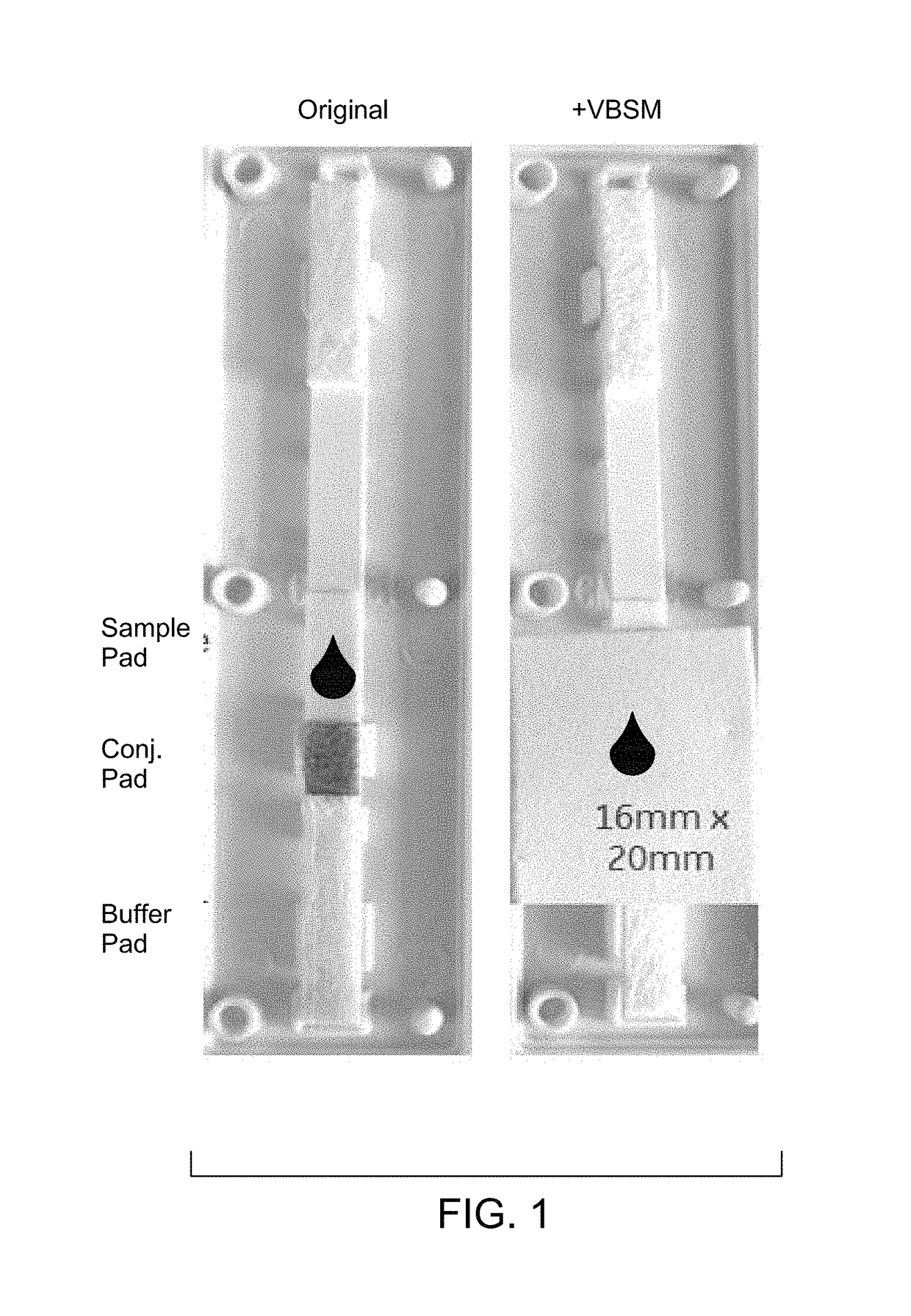
[0071]Materials: CTK HRP2 device RDT (R0114C); Vertical blood separation membrane (VBSM) (asymmetric polysulfone).

[0072]Cut the asymmetric membrane into dimensions of 16 mm×20 mm. Open the CTK housing. Place the membrane on the CTK strip (shiny side facing down), covering sample, conjugate, and part of buffer pads as shown in FIG. 1. Close the CTK housing, or leave it open if being tested shortly.

2) CareStart

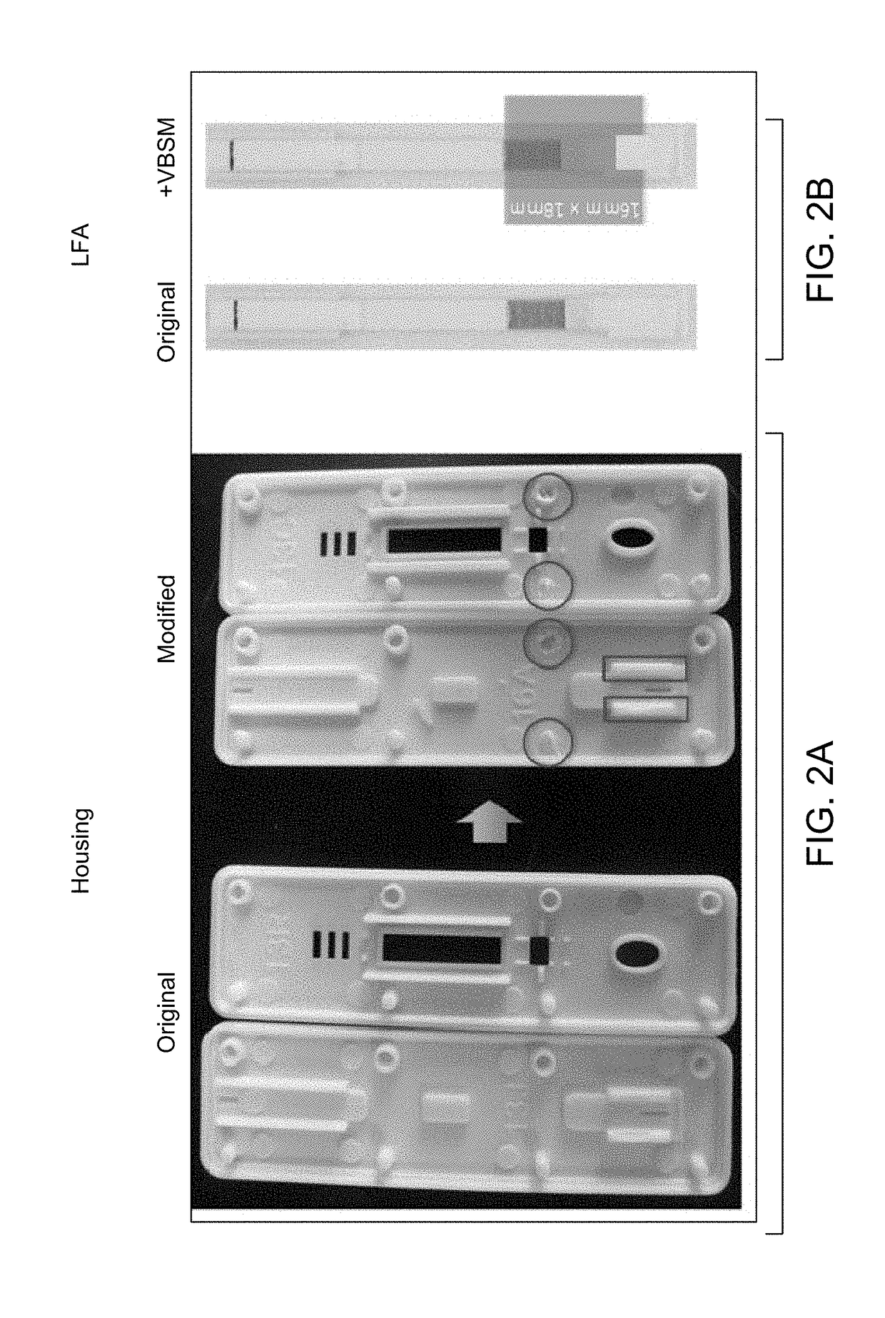
[0073]Materials: CareStart device; Vertical blood separation membrane (VBSM) (asymmetric polysulfone).

[0074]Cut the asymmetric membrane into dimensions of 16 mm×18 mm; Open CareStart housing; Modify housing: remove the second pair of pin point and hole from both the top and bottom plastics, as circled in FIG. 2A; Carefully peel off the buffer pad from conjugate pad, without completely detaching the backing; cut off 5 mm from the upper position. Place VBSM on top of LFA (shiny side facing down), covering conjugate pad, and lay the...

example 2

on of Lateral Channeled Construct with RDTs

1) CTK / SD Bioline

[0083]Materials: CTK / SD Bioline device; lateral blood separation membrane (LBSM) (LF1 from GE).

[0084]Cut the membrane into dimensions of 40 mm×10 mm (L-shape); Open the housing; Place LBSM on the side of the LFA test strip and the leg (10 mm) of the LF1 material touching the middle of conjugate pad, and part of buffer pads (FIG. 7); Close the housing.

Test Procedure

[0085]HRP2 stock aliquots was stored under −80° C.; Prepare HRP2 dilutions in CPD human whole blood at desired concentrations, and store on ice;

[0086]For modified RDTs: Add 75 ul blood sample onto the LBSM at the far end. Allow time for plasma to flow to the conjugate pad and blood to remain on the IBSM. For larger sample volumes, scale LBSM accordingly. Chase with 100 ul non-lytic buffer through buffer port. Cover RDT to minimize evaporation from nitrocellulose. Wait and read results after 20 minutes.

[0087]For regular RDTs as control: Add 5 ul blood sample throug...

example 3

Sensitivity of Integrated Devices

[0089]Materials: CTK HRP2 device RDT (R0114C); Experimental cellulose nanobeads (CNB) device for HRP2; Vertical blood separation membrane (VBSM) (asymmetric polysulfone).

[0090]In this configuration, the original CTK conjugate particle pad is replaced with a cellulose nanobead conjugate pad, and the housing remains the same as original CTK.

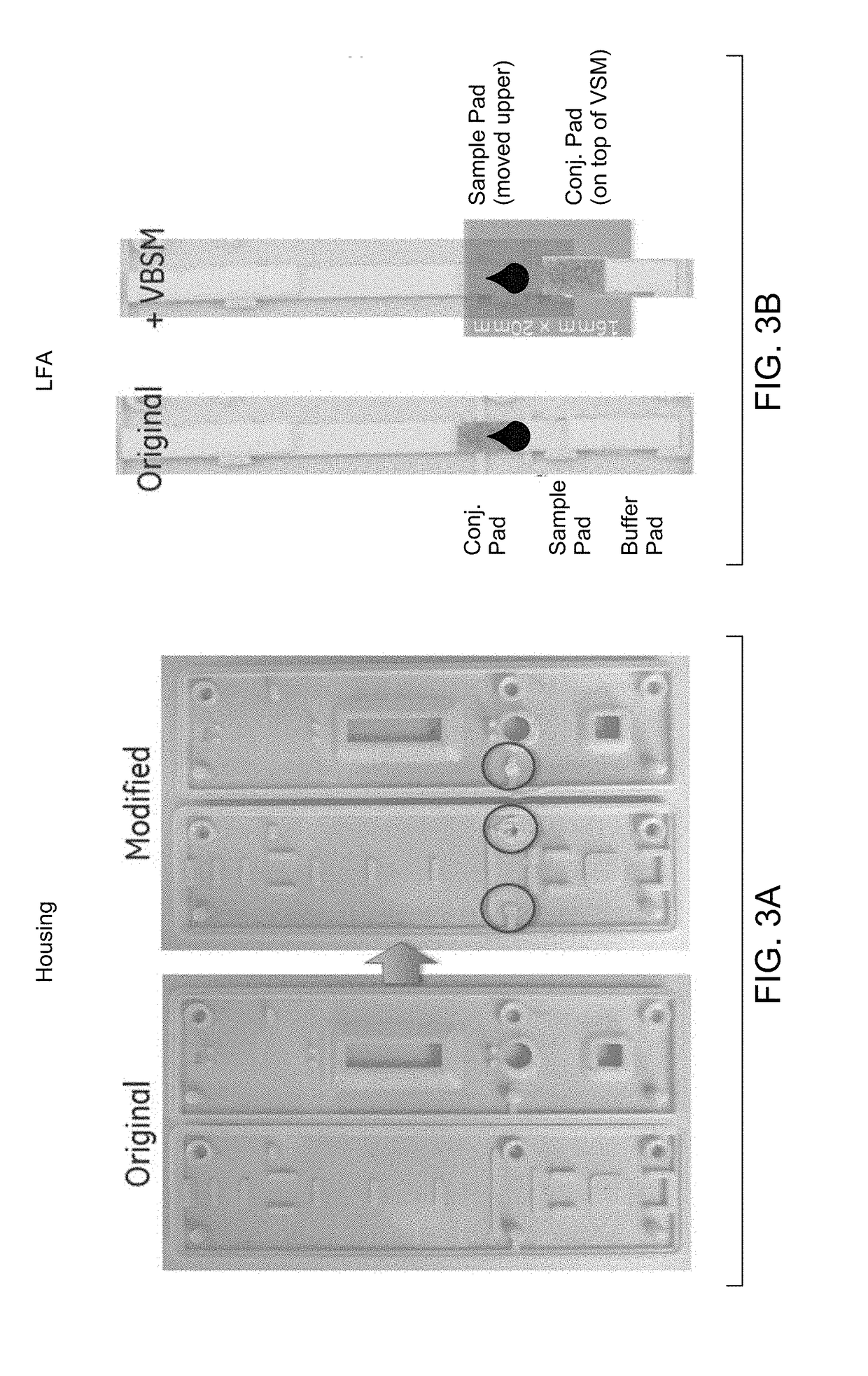
[0091]Cut VBSM into dimensions of 16 mm×20 mm; Open CTK housing. Take off conjugate pad. Replace CTK conjugate with cellulose nanobeads pad. Place VBSM on top of the modified CTK LFA test strip (shiny side facing down), covering sample, conjugate, and part of buffer pads. Close the CTK housing, or leave it open if being tested shortly.

Test Procedure

[0092]Prepare HRP2 (CTK A3005) dilutions in CPD human whole blood (BioReclamation). Open up the housing, add 75 μL blood sample onto center point of the VBSM slowly. Close the housing, and chase with 100 μL non-lytic buffer through buffer port.

[0093]Non lytic buffer 1, fo...


PUM

PropertyMeasurementUnit
volumeaaaaaaaaaa
pore sizeaaaaaaaaaa
pore sizeaaaaaaaaaa

Abstract

Provided herein are integrated devices that improve sensitivity of rapid diagnostic tests. The devices described herein integrate sample separation with rapid diagnostic tests and improve signal intensity of the rapid diagnostic tests.

Description

BACKGROUND[0001]This disclosure relates generally to an improvement in currently available rapid diagnostic tests (RDTs), particularly RDTs for malaria.[0002]A rapid diagnostic test (RDT) is a medical diagnostic test that is quick and easy to perform. RDTs are suitable for preliminary or emergency medical screening, for use in medical facilities with limited resources, and offer a useful alternative to microscopy in situations where reliable microscopic diagnosis is not available or is not available right away. They also allow point-of-care (POC) testing in primary care in situations where formerly only a laboratory test could provide a diagnosis. RDTs do not require clinical diagnostic methods, such as enzyme-linked immunosorbent assay (ELISA) or polymerase chain reaction (PCR), can be performed independent of laboratory equipment by minimally trained personnel, and deliver instant results. RDTs provide results within two hours, and typically provide results in approximately 30 min...

Claims


Application Information

Patent Timeline
Patent Type & Authority Applications(United States)
IPC IPC(8): G01N33/569
CPCG01N2333/445G01N33/56905G01N33/54393G01N33/54388
Inventor LI, BINGMISNER, MATTHEW JEREMIAHGROSSMANN, GREGORY ANDREWOLSEN, CATHRYN ELLENNATARAJAN, ARUNKUMAR
Owner GENERAL ELECTRIC CO
Why Eureka
Try Eureka