Laser-assisted hydrogel microneedle array biomarker extraction and detection device and preparation method thereof

A biomarker and microneedle array technology, which is applied in the field of laser-assisted hydrogel microneedle array extraction and detection of biomarkers in blood devices and its preparation, can solve the problems of damaged capillaries, low concentration of biomarkers, and the detection of analytes. Insufficient extraction volume and other problems, to achieve the effect of improving detection sensitivity, reliable extraction and detection, and excellent biological safety

Pending Publication Date: 2019-12-10
SUN YAT SEN UNIV
View PDF9 Cites 6 Cited by
  • Summary
  • Abstract
  • Description
  • Claims
  • Application Information

AI Technical Summary

Problems solved by technology

However, the use of microneedle arrays to extract subcutaneous biomarkers often encounters the problem of insufficient extraction of analytes, such as: the concentration of most biomarkers in the dermis is too low, so that microneedle arrays cannot capture them and carry out detection
To overcome this defect, the length of the microneedle needs to be increased to penetrate to the lower layer of the dermis, but the dermis contains nerves and capillaries. When the microneedle touches the nerves and capillaries, it will cause pain and bleeding for the user, reducing the user's sensitivity to the microneedle. Compliance with detection methods, there is also a risk of infection
In addition, there was large variation between individual microneedles in the same array, mainly because overly long microneedles penetrated into the subdermis, damaging capillaries and allowing biomarker leakage
Only a few microneedles in each array disrupted capillaries, caused local bleeding and showed strong binding ability of local microneedles to biomarkers, while most of the remaining microneedles in the array could only capture biomarkers in interstitial fluid. A small amount of biomarkers may not be captured. The low uniformity of microneedle array capture makes it difficult to quantify biomarkers in an array, and it is impossible to stably detect a single biomarker. Therefore, The sensitivity of microneedle array detection technology still needs to be improved, and it is difficult to apply it in actual diagnosis at present

Method used


Image

  • Laser-assisted hydrogel microneedle array biomarker extraction and detection device and preparation method thereof
  • Laser-assisted hydrogel microneedle array biomarker extraction and detection device and preparation method thereof
  • Laser-assisted hydrogel microneedle array biomarker extraction and detection device and preparation method thereof

Examples


Embodiment Construction

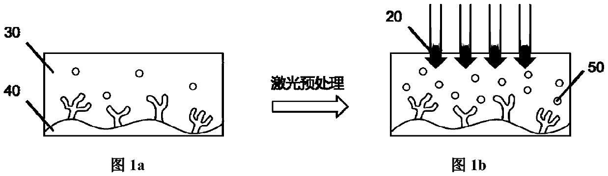
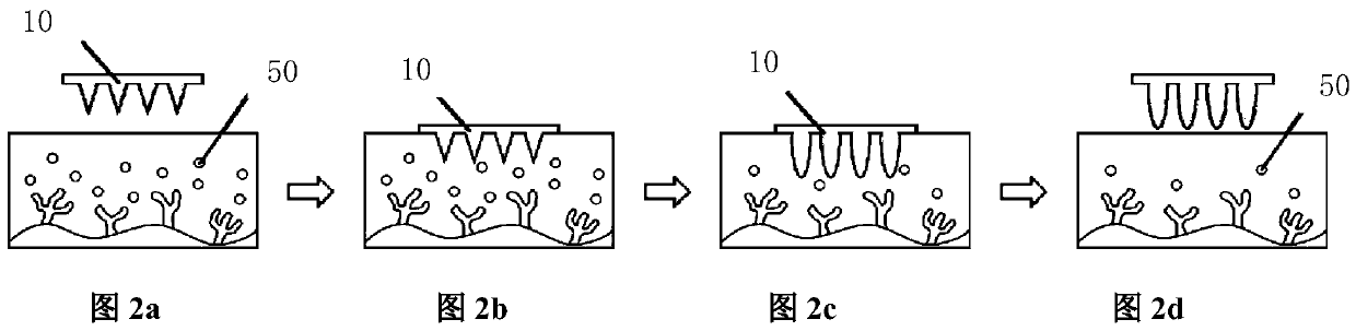
[0044] The laser-assisted hydrogel microneedle array extraction and detection device for biomarkers in blood according to the present invention includes a hydrogel microneedle array 10 and a laser irradiation device 20, and the hydrogel microneedle array 10 is composed of solute mass ratio The range value is 2:1 to 10:1 perfluoromethyl vinyl ether (PMVE) aqueous solution and polyethylene glycol (PEG, M W =10000) aqueous solution, for example, can choose mass fraction 11.2% perfluoromethyl vinyl ether (PMVE) aqueous solution and mass fraction 5.6% polyethylene glycol (PEG, M W =10000), the wavelength range value of the laser irradiation device 10 is 530-600nm.

[0045] Such as figure 1 a- figure 1 b. figure 2 a-2d and image 3 , Figure 4 As shown, the skin 30 is pretreated to increase the permeability of the subcutaneous blood vessels 40 to the biomarkers 50 in the blood, so that the biomarkers 50 in the subcutaneous blood at the irradiation site are enriched in the subc...


PUM


Abstract

The invention belongs to the technical field of medical examination, and particularly discloses a laser-assisted hydrogel microneedle array biomarker extraction and detection device which comprises ahydrogel microneedle array and a laser irradiation device, and biomarkers are extracted through the hydrogel microneedle array after irradiation of the laser irradiation device. The device can be usedfor efficiently extracting the biomarker; by means of the characteristics of water absorption and swelling of the hydrogel microneedle array, the device can effectively extract enough biomarkers fromtissue fluid, has excellent biological safety to skin, does not influence normal functions of human skin, extracts and detects the biomarkers under the corium layer, and is painless, minimally invasive and capable of rapidly detecting diseases.

Description

technical field [0001] The invention belongs to the technical field of medical examination for extracting and detecting biomarkers in blood, in particular to a device for extracting and detecting biomarkers in blood with laser-assisted hydrogel microneedle arrays and a preparation method thereof. Background technique [0002] In the prior art, blood biochemical routine examination is a common test item in medical examination, and it is also an important way to discover potential diseases. Among them, the test items reflect a number of important indicators in the function of human organs: blood lipids, Blood sugar, kidney function, liver function, etc. Many diseases have no typical symptoms in the early stage of the disease, but the corresponding biomarkers of the disease are abnormal, and blood tests are required to detect them. For example, the early stage of diabetes is a typical case, and the early stage is manifested as abnormal blood sugar, and the early stage of gout i...

Claims


Application Information

Patent Timeline
Patent Type & Authority Applications(China)
IPC IPC(8): G01N1/28G01N21/64
CPCG01N1/28G01N21/6402G01N21/6458G01N21/6486
Inventor 谢曦黄新烁陈惠琄杭天胡宁何根李湘凌杨成端
Owner SUN YAT SEN UNIV
Why Eureka
Try Eureka