Eureka AIR delivers breakthrough ideas for toughest innovation challenges, trusted by R&D personnel around the world.

Method for predicting complications of normal tissue organs after tumor radiotherapy

A radiotherapy technology for tissues, organs and tumors, applied in radiotherapy, X-ray/γ-ray/particle irradiation therapy, treatment, etc., can solve the problems of not being widely applicable, lack of pertinence and planning, and low degree of accuracy, and achieve improvement Effect of treatment and quality of life in the later stage, improvement of prediction accuracy, effect of reducing side effects and complications

Active Publication Date: 2019-12-24
CANCER CENT OF GUANGZHOU MEDICAL UNIV
View PDF11 Cites 9 Cited by
  • Summary
  • Abstract
  • Description
  • Claims
  • Application Information

AI Technical Summary

Problems solved by technology

Studies have shown that the application of radiomics data can predict the complications of tumor patients after radiotherapy and chemotherapy, so as to provide timely and effective treatment and intervention for patients, and reduce the occurrence of complications. Insufficient sex leads to low accuracy and is still not widely applicable. Therefore, an efficient and orderly prediction method for patients is needed to improve the prediction accuracy, which is of great significance for improving the treatment effect of patients and the quality of life in the later period.

Method used


Image

  • Method for predicting complications of normal tissue organs after tumor radiotherapy

Examples


specific Embodiment approach

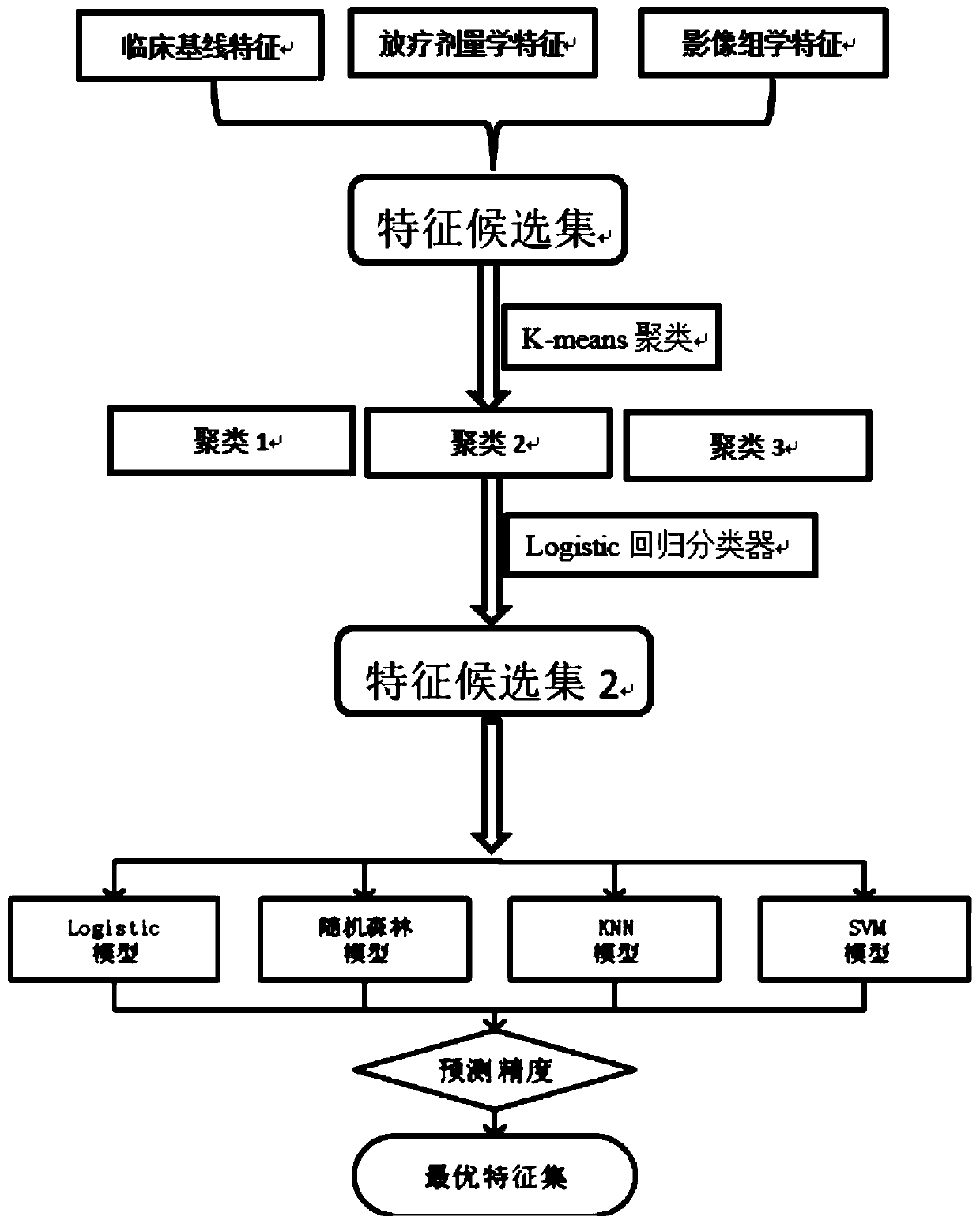
[0033] A method for predicting complications of normal tissues and organs after radiotherapy for tumors, characterized in that the method for predicting mainly includes the following steps:

[0034] S1. Obtain Y modal image data of several radiotherapy patients in X periods, and obtain information on the occurrence, type, and time of organ-at-risk complications of radiotherapy patients after radiotherapy, for example, the time of occurrence is after the end of treatment In the mth month, a multimodal image database is established; further, multimodal image data include diagnostic CT, simulated positioning CT, different sequences of multi-parameter MR images, PET / CT, B-ultrasound images, CBCT images and conventional X-rays image data;

[0035] S2. Using a segmentation method to extract image data of organs at risk near the tumor target area from the multimodal image database; the segmentation method is automatic or manual segmentation, and automatic segmentation includes but is...


PUM


Abstract

The invention relates to a technology for predicting clinical complications after radiotherapy of tumor patients, in particular to a method for predicting complications of normal tissue organs after tumor radiotherapy based on the multimodal image omics characteristics and the radiotherapy dosimetry characteristics. The method mainly comprises the steps: S1, a multimodal image database is established; S2, image data of endangered organs near a tumor target area are extracted; S3, the image omics characteristics of the endangered organs are extracted, and the characteristics of normal organ image data are extracted; S4, parameters of the image phenotypic characteristics of the endangered organs are extracted according to image segmentation results; S5, the image omics characteristics are analyzed; S6, parameters of the exposure doses of the endangered organs are extracted; and S7, the characteristics are collected and extracted. The complications after radiotherapy and chemotherapy of the tumor patients are predicted through the image omics data, effective treatment and intervention can be provided for the patients in time through the reliable, safe and high-precision prediction method, the complications are reduced, and thus the treatment effect on the patients and the later life quality of the patients are improved.

Description

technical field [0001] The invention relates to a technique for predicting clinical complications after radiotherapy for tumor patients, in particular to a method for predicting complications of normal tissues and organs after radiotherapy for tumors based on multimodal radiomics features and radiotherapy dosimetry features. Background technique [0002] Malignant tumors are major diseases that seriously threaten human health. Radiotherapy and chemotherapy are two commonly used methods in the treatment of many cancer patients. Considering the possibility of postoperative complications in cancer patients, the target area should be increased as much as possible during radiotherapy and chemotherapy. (Tumor) dose while reducing radiation damage to normal tissues (organs at risk) around the target area is the ultimate goal of radiation therapy. [0003] Since the location of the tumor is usually located in the patient's body, the radiation must pass through some normal tissues wh...

Claims


Application Information

Patent Timeline
Patent Type & Authority Applications(China)
IPC IPC(8): A61B5/00A61N5/10
CPCA61B5/7275A61B5/7267A61N5/1031A61N5/1071
Inventor 张国前张书旭谭剑明余辉王琳婧彭莹莹周露王锐濠张全彬阳露雷怀宇沈国辉廖煜良李萍曾庆星
Owner CANCER CENT OF GUANGZHOU MEDICAL UNIV
Why Eureka
Eureka Blog
Learn More