Chemiluminescence biosensor for detecting uracil-DNA glycosylase and preparation method and application of chemiluminescence biosensor

A uracil glycosylase and biosensor technology, which is applied in the field of biosensors to achieve the effects of mild reaction conditions, improved efficiency and highly sensitive detection

Active Publication Date: 2019-12-24
UNIV OF JINAN
View PDF6 Cites 1 Cited by
  • Summary
  • Abstract
  • Description
  • Claims
  • Application Information

AI Technical Summary

Problems solved by technology

[0004] Aiming at the current lack of an efficient, sensitive and convenient method for detecting uracil glycosylase, the present invention provides a chemiluminescence technology for detecting uracil based on three-way structure-driven strand displacement reaction and DNA walker technology to drive spherical nuclease Glycosylase mainly includes the construction of two nanostructures of three-way structure and spherical nucleic acid, using

Method used


Image

  • Chemiluminescence biosensor for detecting uracil-DNA glycosylase and preparation method and application of chemiluminescence biosensor
  • Chemiluminescence biosensor for detecting uracil-DNA glycosylase and preparation method and application of chemiluminescence biosensor
  • Chemiluminescence biosensor for detecting uracil-DNA glycosylase and preparation method and application of chemiluminescence biosensor

Examples


preparation example Construction

[0051] The preparation method of described biosensor comprises the following steps:

[0052] (1) Preparation of gold nanoparticles;

[0053] (2) Preparation of spherical nucleic acid;

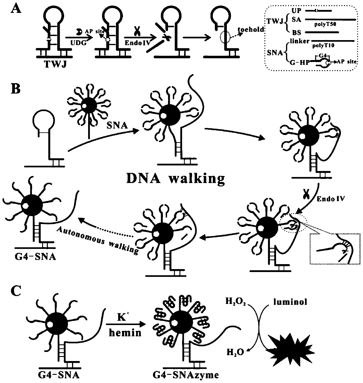
[0054] (3) The formation of spherical nuclease in homogeneous phase is used to catalyze the chemiluminescent reaction of luminol;

[0055] In the described preparation method, the preparation of nano gold:

[0056] The preparation of nano-gold is realized by reducing chloroauric acid with sodium citrate. Add 500μL of chloroauric acid (0.04g / mL) into 200mL of ultrapure water, stir and heat until boiling, then add 3mL of sodium citrate (1%) and quickly add to the boiling solution. Subsequently, it can be observed that the color of the solution changes from light yellow to black and finally to wine red. Continue heating for 15 min after turning wine red to ensure complete reaction. Afterwards, the nano-gold solution was cooled to room temperature and placed at 4°C for use. The UV absorption ...

Embodiment 1

[0058] Preparation of spherical nucleic acids:

[0059] First, the nano-gold stock solution was centrifuged at 13000r / min and 4°C for 20min, then the supernatant was removed and the bottom precipitate was dispersed in ultrapure water to make the concentration 5nM. Then, 150 μL of 10 μM DNA strand (V G-HP:Vlinker =20:1) was added to the nano-gold mixture, and left for 24 hours (4°C) after the addition. Afterwards, 50 μL of PB buffer (10 mM PB, pH 7.4) and 27 μL of PBS (10 mM PB, 2M NaCl, pH 7.4) were added to the mixture. After 48 hours (4°C), continue to add 62 μL of PBS, at this time the concentration of NaCl in the solution is 0.3M. After 24 hours, the mixture was eluted three times by centrifugation at 13000 r / min for 15 minutes to remove unlabeled DNA strands. Finally, the centrifuged precipitate was redissolved in 100 μL of ultrapure water and kept at 4°C for use.

[0060] The spherical nucleic acid has been prepared so far, and the main steps of the reaction process ...

Embodiment 2

[0064] Preparation of spherical nucleic acids:

[0065] First, the nano-gold stock solution was centrifuged at 13000r / min and 4°C for 20min, then the supernatant was removed and the bottom precipitate was dispersed in ultrapure water to make the concentration 5nM. Then, 150 μL of 10 μM DNA strand (V G-HP:Vlinker =20:1) was added to the nano-gold mixture, and left for 24 hours (4°C) after the addition. Afterwards, 50 μL of PB buffer (10 mM PB, pH 7.4) and 27 μL of PBS (10 mM PB, 2M NaCl, pH 7.4) were added to the mixture. After 48 hours (4°C), continue to add 62 μL of PBS, at this time the concentration of NaCl in the solution is 0.3M. After 24 hours, the mixture was centrifuged at 13,000 r / min for 15 min and eluted three times to remove unlabeled DNA strands. Finally, the centrifuged precipitate was redissolved in 100 μL of ultrapure water and kept at 4°C for use.

[0066] The spherical nucleic acid has been prepared so far, and the main steps of the reaction process in th...


PUM

PropertyMeasurementUnit
Extinction coefficientaaaaaaaaaa

Abstract

The invention relates to the technical field of a biosensor, in particular to detection of uracil-DNA glycosylase based on a chemiluminescence technique for driving a strand displacement reaction based on a tee structure and driving spherical nucleases based on a DNA walker technique. In order to solve the problems of being complex to operate and low in sensitivity for a method for detecting uracil-DNA glycosylase in the prior art, the invention provides a biosensor based on two nanometer technologies of the tee structure and DNA walker, and spherical nucleases are used for catalyzing luminalto be subjected to a chemiluminescence reaction for detection. The preparation method comprises the steps of preparing nano-Au, preparing spherical nucleic acid, and forming spherical nucleases in homogeneous phase for catalyzing the chemiluminescence reaction of the luminal. The uracil-DNA glycosylase is used for performing specific recognition and excision on U alkali, so as to realize target specificity detection. Besides, the DNA walker nanometer technology is used for realizing quick and high-sensitivity detection on targets.

Description

technical field [0001] The invention relates to the technical field of biosensors, in particular to a chemiluminescent biosensor for detecting uracil glycosylase based on a three-way structure and DNA walker nanotechnology and a preparation method thereof, and also to spherical nuclease technology. Background technique [0002] The integrity of DNA is crucial to maintaining the function of organisms. When DNA is damaged spontaneously, the base excision repair mechanism (BER) will be activated to repair DNA damage. The BER repair pathway is mainly initiated by DNA glycosylase, in which uracil glycosylase (UDG) specifically recognizes and removes uracil U formed by deamination of cytosine in DNA to form an AP site. Under the action of apurine / pyrimidine endonuclease 1 (APE1) or endonuclease IV (Endo IV), the DNA chain is cut to generate a chain break, and the DNA chain is repaired through the joint action of subsequent DNA polymerase and ligase. Uracil glycosylase plays a ver...

Claims


Application Information

Patent Timeline
IPC IPC(8): C12Q1/48G01N21/76
CPCC12Q1/48G01N21/76
Inventor 王玉张雪刘素黄加栋宋晓蕾李莎莎王敬锋王海旺孙文玉王业茹
Owner UNIV OF JINAN
Why Eureka
Try Eureka