Hip joint segmentation method and device, electronic equipment and storage medium
A hip joint and segmentation model technology, applied in the field of medical image processing, can solve problems such as cumbersome operation, time-consuming operation, and high technical difficulty, and achieve the effect of meeting clinical needs and obtaining accurate results
- Summary
- Abstract
- Description
- Claims
- Application Information
AI Technical Summary
Problems solved by technology
Method used
Image
Examples
Embodiment 1
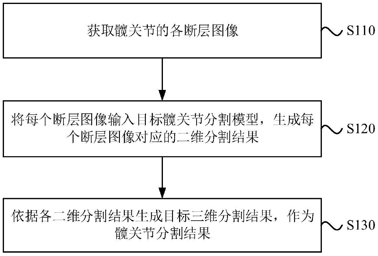
[0030] The hip joint segmentation method provided in this embodiment can be applied to the hip joint bone structure segmentation and hip joint classification of medical images. The method can be executed by a hip joint segmentation device, which can be implemented by software and / or hardware, and the device can be integrated in a device with image processing functions, such as a personal computer, a server, or a network device. See figure 1 , The method of this embodiment specifically includes the following steps:
[0031] S110. Obtain various tomographic images of the hip joint.
[0032] In order to obtain the three-dimensional segmentation result of the hip joint of the scanned object, a three-dimensional image containing the hip joint of the scanned object needs to be obtained first. The three-dimensional image may be in a three-dimensional form or may be a stack of multiple two-dimensional images (tomographic images). Since the hip joint segmentation directly based on the thre...
Embodiment 2
[0051] This embodiment adds training related steps of the "target hip joint segmentation model" on the basis of the first embodiment above. The explanation of the terms that are the same as or corresponding to the above-mentioned embodiments will not be repeated here.
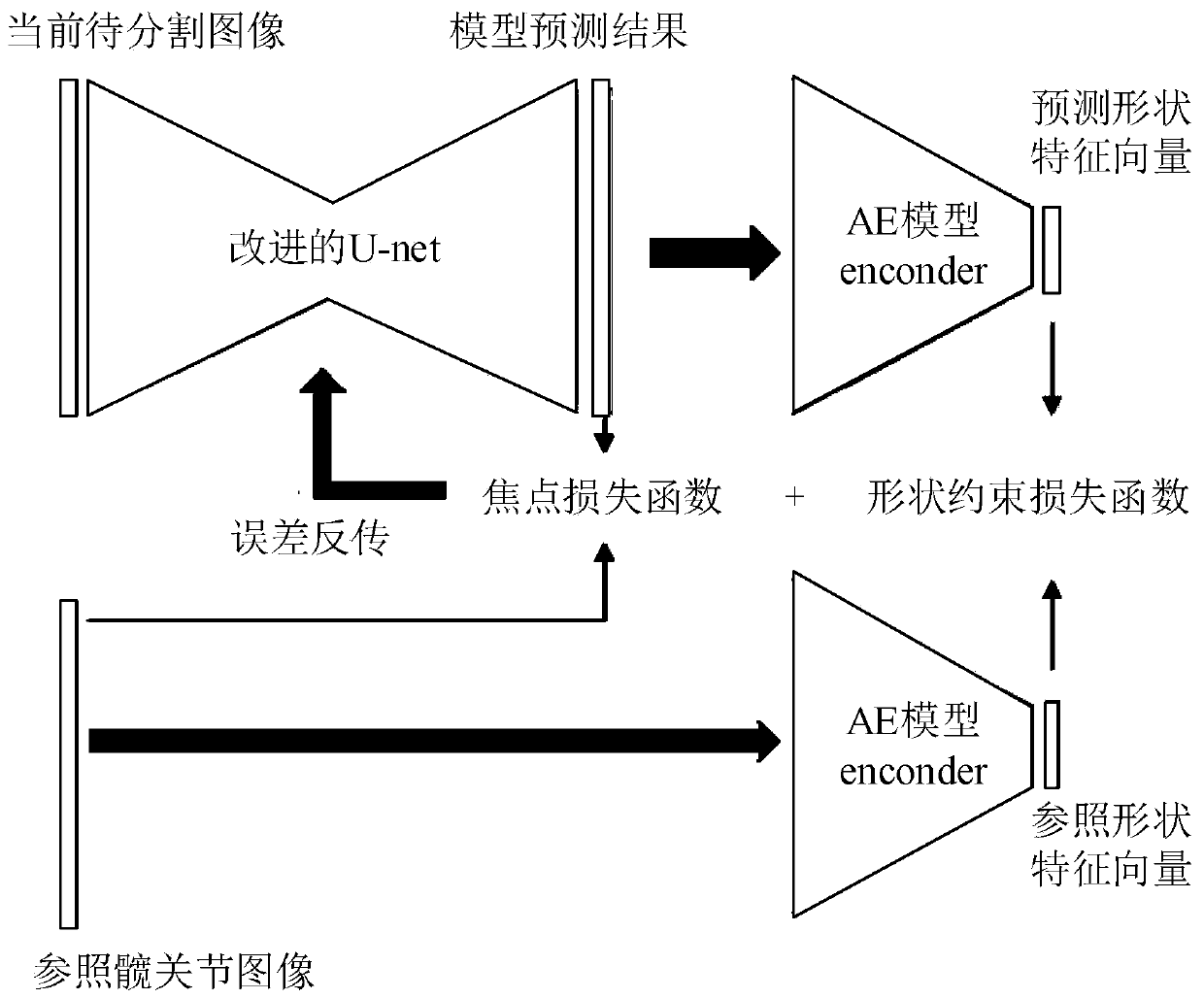
[0052] In this embodiment, before describing the training method of the target hip joint segmentation model, the model structure of the target hip joint segmentation model is described first.
[0053] Exemplarily, the basic model of the target hip joint segmentation model is a full convolutional neural network model based on two-dimensional medical images, and the loss function of the target hip joint segmentation model is a focus loss function and a shape constraint loss function; wherein, the shape constraint loss The function is the Euclidean distance between the shape feature vector in the model prediction result and the shape feature vector in the reference hip joint image corresponding to the current image to ...
Embodiment 3
[0077] This embodiment provides a hip joint segmentation device, see Image 6 , The device specifically includes:
[0078] The tomographic image acquisition module 610 is used to acquire various tomographic images of the hip joint;
[0079] The two-dimensional segmentation result generation module 620 is configured to input each tomographic image into the target hip joint segmentation model to generate a two-dimensional segmentation result corresponding to each tomographic image, wherein the target hip joint segmentation model is pre-trained based on the convolutional neural network model obtain;
[0080] The hip joint segmentation result generation module 630 is configured to generate a target three-dimensional segmentation result according to each two-dimensional segmentation result as the hip joint segmentation result.
[0081] Optionally, the two-dimensional segmentation result generating module 620 is specifically configured to:
[0082] For each tomographic image, according to th...
PUM
 Abstract
Description
Claims
Application Information
 - R&D
- Intellectual Property
- Life Sciences
- Materials
- Tech Scout
- Unparalleled Data Quality
- Higher Quality Content
- 60% Fewer Hallucinations
Browse by: Latest US Patents, China's latest patents, Technical Efficacy Thesaurus, Application Domain, Technology Topic, Popular Technical Reports.
© 2025 PatSnap. All rights reserved.Legal|Privacy policy|Modern Slavery Act Transparency Statement|Sitemap|About US| Contact US: help@patsnap.com


