
Spinal endoscope hemostatic forceps

A technology of hemostatic forceps and spine, applied in the field of medical devices, can solve the problems of increased local infection rate of patients, difficult to heal injuries, nerve dysfunction and other problems, and achieve the effect of reducing peripheral control components, avoiding deterioration, and reducing space occupancy

Pending Publication Date: 2020-01-14
CHONGQING TRADITIONAL CHINESE MEDICINE HOSPITAL
View PDF0 Cites 0 Cited by
  • Summary
  • Abstract
  • Description
  • Claims
  • Application Information

AI Technical Summary

Problems solved by technology

[0004] The inventor, as the chief surgeon of the pain department, found the following drawbacks in the prior art during the long-term treatment of patients. When the patient is bleeding, the endoscope vision is blurred and bone wax cannot be used to stop the bleeding. Therefore, medical cotton must be used to stop the bleeding first. At this time, the patient lies on the operating bed for a long time, which will increase the local infection rate of the patient, increase the nerve compression, and even lead to postoperative neurological dysfunction or even disability, prolonging the pain of the patient's operation. Unable to effectively stop bleeding, minimally invasive surgery is usually converted into open surgery (at this time, local anesthesia becomes general anesthesia), and at the same time, the damage to the patient is increased, and even these injuries are difficult to heal
Even though the medical cotton can reduce the patient's blood loss and regain the vision of the medical staff, the bone wax needs the medical staff to knead and shape it. At the same time, the bone wax tends to stick to the endoscope during transmission, causing the lost vision again.

Method used


Image

  • Spinal endoscope hemostatic forceps
  • Spinal endoscope hemostatic forceps
  • Spinal endoscope hemostatic forceps

Examples


Embodiment 1

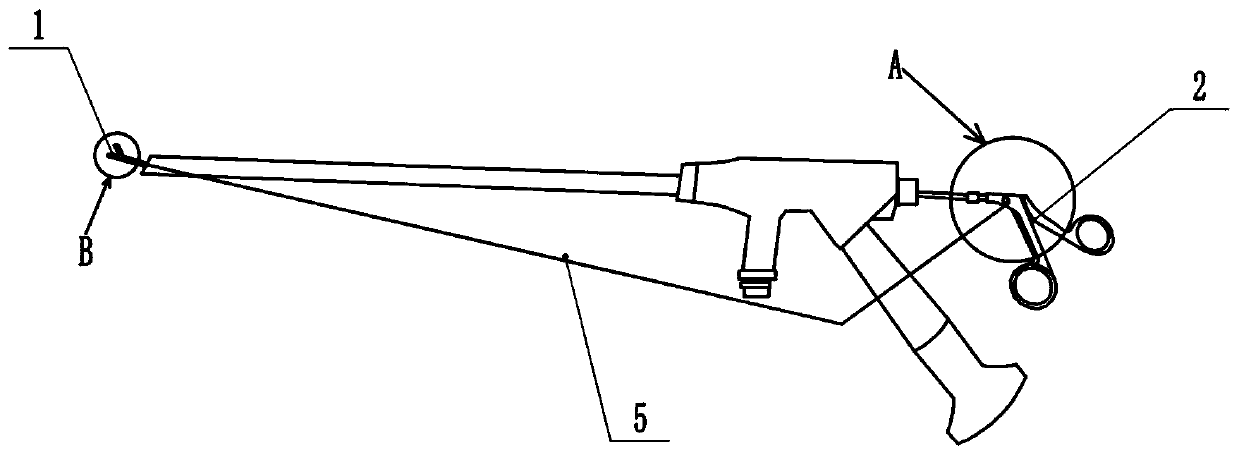
[0038] Basic as attached figure 1 As shown: a spinal endoscopic hemostat includes a clamping end that contacts the working surface and a handle 2 for controlling the clamping end. The clamping end is divided into a working layer 1, a discharge layer 3, and a filling layer 4 from top to bottom. The working layer 1, the discharge layer 3 and the filling layer 4 are spaced apart from each other, and a feed port is opened at the intersection of the discharge layer 3 and the filling layer 4.

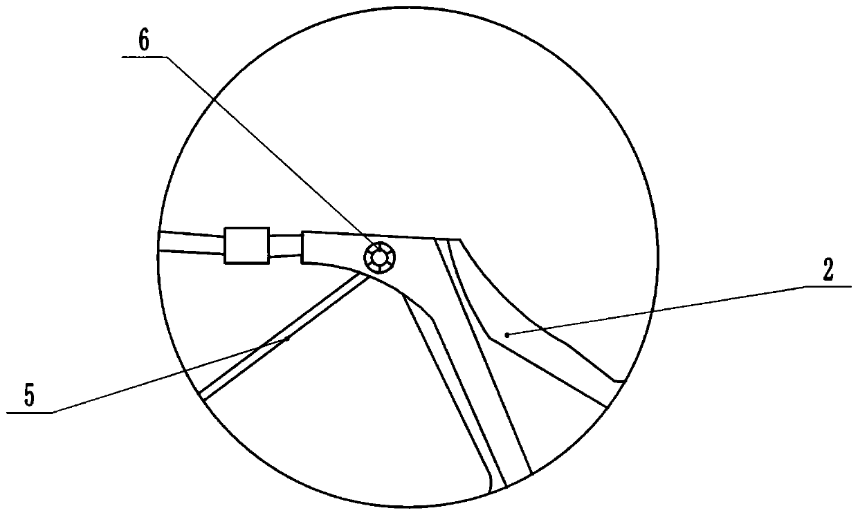
[0039] Please refer to Figure 4 , the discharge layer 3 includes a horizontally placed slide plate 301, an axially placed tension spring 302, a vertical plectrum 303 and a connecting rod 5 hinged to each other, the slide plate 301 is located in the movement stroke of the plectrum 303, and the slide plate 301 covers On the surface of the material supply port, a tension spring 302 placed axially is hinged between the slide plate 301 and the discharge layer 3, and a discharge port 12 is opened...

Embodiment 2

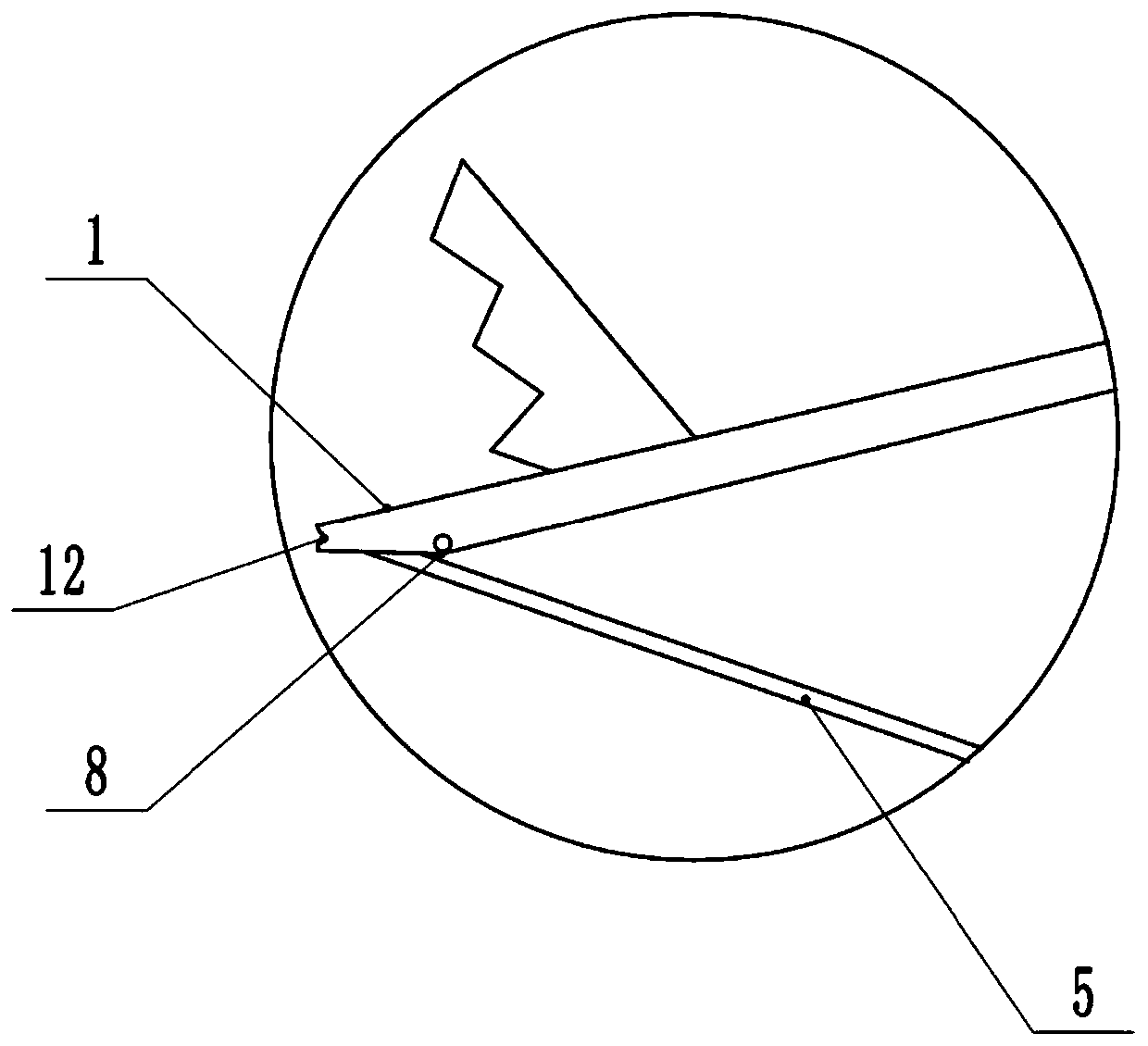
[0051] Please refer to Image 6 with Figure 7 , this embodiment is a simplified solution of Embodiment 1, wherein the difference between Embodiment 2 and Embodiment 1 is that it also includes a manual filling layer, a connecting rod 13 is connected between the clamping end and the handle, and the side of the connecting rod 13 There is a discharge hole on the wall, and a push-fit plate 14 is slidingly connected between the discharge hole and the side wall. A feed plate 15 is provided behind the discharge hole, and a parallel multi-connector for easy extension and contraction is hinged between the feed plate 15 and the handle. The rod device 16 has an outlet 17 with a tapered cross section at the maximum stroke of the feeding plate 15 .

[0052] The specific implementation is as follows. When performing bone wax hemostasis, in addition to the method in Embodiment 1, the medical staff can also open the push plate 14 of the connecting rod 13, put the bone wax into the discharge ...


PUM


Abstract

The invention discloses a pair of spinal endoscope hemostatic forceps in the field of medical instruments, which comprises a forceps clamping end in contact with a working surface and a handle for controlling the forceps clamping end, the forceps clamping end is sequentially divided into a working layer, a discharging layer and a filling layer, the working layer, the discharging layer and the filling layer are spaced from one another, and a feeding hole is formed in the junction of the discharging layer and the filling layer; a working piece for orthopaedic excision is machined on the surfaceof the working layer; a sliding piece, an elastic piece, a pushing piece and a control piece are arranged in the discharging layer; the sliding piece is located in a movement stroke of the pushing piece. The sliding part covers the surface of the feeding opening; an elastic piece is hinged between the sliding piece and the discharging layer; a discharging opening is formed in the maximum stroke position of the pushing piece and located in the end opening of the discharging layer, a control piece for controlling the pushing piece to reciprocate is connected between the pushing piece and the handle, the medical staff operate the pushing piece to convey bone wax to the discharging opening, bleeding stopping is conducted on the bone bleeding position of a patient in time, the operation difficulty is lowered, and bone wax conveying time is shortened.

Description

technical field [0001] The invention belongs to the field of medical instruments, in particular to a spinal endoscopic hemostatic forceps. Background technique [0002] In the 1980s, with the development of technology and endoscopic instruments, orthopedic surgeons began to use endoscopic technology for spinal surgery. After the 1990s, endoscopic spinal surgery has made great progress and is currently more clinically practical. Valuable techniques include endoscopic assisted posterior or lateral posterior approach of intervertebral discectomy, laparoscopy assisted lumbar debridement, and thoracoscopic assisted thoracic debridement. [0003] When performing minimally invasive orthopedic or pain surgery, occasional bone bleeding may occur in patients. In the prior art, when performing hemostasis in minimally invasive surgery, the medical staff usually use the following techniques. When blood appears in the endoscopic field of view (more than 2ml), the medical staff usually us...

Claims


Application Information

Patent Timeline
IPC IPC(8): A61B17/12A61B17/28
CPCA61B17/12A61B17/282A61B17/2833A61B17/2841A61B2017/12004
Inventor 谈俊廖翔
Owner CHONGQING TRADITIONAL CHINESE MEDICINE HOSPITAL
Learn More