
Cell nucleus DNA dyeing method

A dyeing method and cell nucleus technology, applied in sampling, measuring devices, instruments, etc., can solve problems such as shelf life hindering reagent storage, acid hydrolysis time and temperature influence, complex preparation, etc., to achieve shortened dyeing time, less human stimulation, and simple preparation Effect

Inactive Publication Date: 2020-01-31
MOTIC XIAMEN MEDICAL DIAGNOSTICS SYST
View PDF2 Cites 1 Cited by
  • Summary
  • Abstract
  • Description
  • Claims
  • Application Information

AI Technical Summary

Problems solved by technology

Due to the cumbersome staining process and long staining time of the traditional method, it is easily affected by the type of tissue fixative, acid hydrolysis time and temperature, which directly affects the accuracy of the measured value of DNA content in cells.
The hydrochloric acid used in the traditional method is a precursor chemical, which is strictly controlled and difficult to purchase, and its volatility is not conducive to human health
The preparation of traditional cell DNA staining solution is complicated, boiling is required during the process, and the effective storage period is about 2-5 days. Such a short shelf life hinders the storage of this reagent and also hinders its commercial application.

Method used


Image

  • Cell nucleus DNA dyeing method
  • Cell nucleus DNA dyeing method
  • Cell nucleus DNA dyeing method

Examples


Embodiment 1

[0042] A method for staining cell nucleus DNA, comprising the following steps:

[0043] 1, be 1: 9 formaldehyde solution and 95% ethanol preparation AF fixative according to volume ratio;

[0044] II. Prepare cell DNA staining solution, hydrolyzate, rinsing solution and eosin staining solution according to Table 1;

[0045] Table 1

[0046]

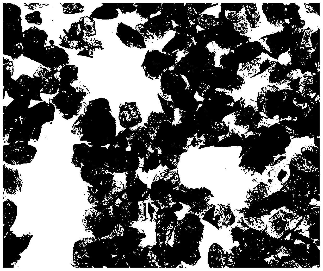
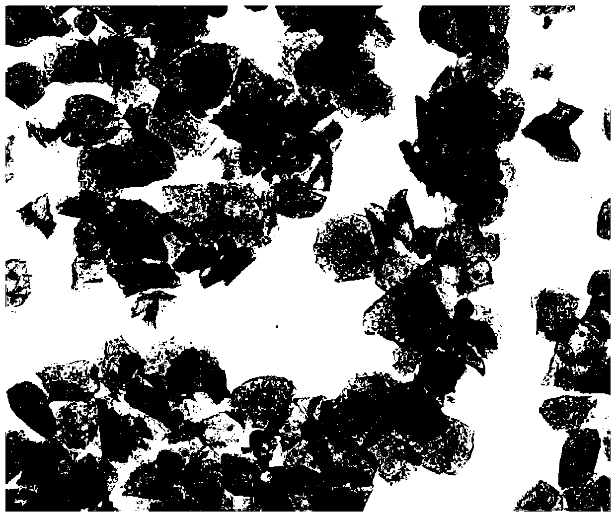
[0047] III. Put the pathological sample in AF fixative solution for 10-15 minutes; after washing with running water, put it into hydrolysis solution for 15-30 minutes; after washing with running water, put it into DNA staining solution for staining for 20-40 minutes; Rinse in rinsing solution for 5-10 minutes; dehydrate step by step with 50% ethanol, 75% ethanol, and 95% ethanol, put in eosin staining solution for 1-3 minutes, decolorize with absolute ethanol, dry in the air, and seal the slides. The effect picture after dyeing is as follows figure 2 shown.

Embodiment 2

[0049] The difference from Example 1 is that cell DNA staining solution, hydrolysis solution, rinsing solution and eosin staining solution were prepared according to Table 2.

[0050] Table 2

[0051]

[0052] The effect picture after dyeing is as follows image 3 shown.

Embodiment 3

[0054] The difference from Example 1 is that cell DNA staining solution, hydrolysis solution, rinsing solution and eosin staining solution were prepared according to Table 3.

[0055] table 3

[0056]

[0057]

[0058] The effect picture after dyeing is as follows Figure 4 shown.


PUM


Abstract

The invention discloses a cell nucleus DNA staining method. The method comprises the following steps: preparing an AF stationary liquid, a hydrolysate, a rinsing liquid and an eosin staining liquid; preparing a cell DNA staining solution which is composed of a cell DNA staining solution A and a cell DNA staining solution B in a volume ratio of 1: 1; placing a pathological sample into the AF stationary liquid to be fixed; washing the sample with running water, and then placing the sample into a hydrolysate to hydrolyze; washing the sample with running water, and then placing the sample in the cell DNA staining solution for staining; washing the sample with running water, and then placing the sample in a rinsing liquid for rinsing; and carrying out dehydrating with ethanol step by step, placing the sample into an eosin staining solution, and carrying out decolorizing with absolute ethyl alcohol, airing, and sealing. According to the method, dyeing time is greatly shortened, the hydrolysate is composed of non-precursor chemicals and belongs to an environment-friendly reagent, compared with a traditional stationary liquid, the AF stationary liquid does not include acetic acid and lesssimulates the human body, and the cell DNA staining solution is simple to prepare, does not need to be heated and boiled, has the validity period longer than 18 months, is ready-to-use and is suitablefor commercial popularization.

Description

technical field [0001] The invention relates to a staining method, in particular to a cell nucleus DNA staining method. Background technique [0002] Since Van Duijn discovered a thionine-sulfite reagent specially used to stain mouse liver and kidney nuclei in 1954, this technique has been widely used in the determination of nuclear DNA content. Feulgen staining is a staining method that shows the specificity of nuclear DNA. The specific bases in the double-strand DNA can be hydrolyzed by acidic substances, exposing free aldehyde groups (RNA does not have this characteristic), and the free aldehyde groups interact with cells. The blue-purple intermediate products in the DNA staining solution combine to form a blue-purple nucleus, which is the distribution position and exact content of DNA in the nucleus. Due to the cumbersome staining process and long staining time of the traditional method, it is easily affected by the type of tissue fixative, acid hydrolysis time and temp...

Claims


Application Information

Patent Timeline
Patent Type & Authority Applications(China)
IPC IPC(8): G01N1/30
CPCG01N1/30G01N2001/302G01N2001/305
Inventor 黄荣祥余岚岚朱晨雁
Owner MOTIC XIAMEN MEDICAL DIAGNOSTICS SYST
Learn More