Eureka AIR delivers breakthrough ideas for toughest innovation challenges, trusted by R&D personnel around the world.

3D printing semi-pelvic prosthesis

A 3D printing, half pelvis technology, applied in 3D printing, prosthesis, additive manufacturing, etc., can solve the problem that the center of the acetabular cup structure cannot accurately locate the patient's original acetabular center, the hip joint function, and the weight of the prosthesis cannot be obtained. Increase the long-term stability, increase the probability of bone ingrowth, and overcome the loosening of the prosthesis

Inactive Publication Date: 2020-02-21
FOURTH MILITARY MEDICAL UNIVERSITY
View PDF5 Cites 3 Cited by
  • Summary
  • Abstract
  • Description
  • Claims
  • Application Information

AI Technical Summary

Problems solved by technology

However, if the individualized prosthesis is completely customized according to the pelvic defect caused by surgical resection, the problem of higher infection rate after implantation of a larger-volume prosthesis will also occur with the increase of the resection area, and the prosthesis’s weight will increase
At the same time, in the face of complex pelvic structures, objectively, the number of screws required to fix the prosthesis is also large, but in fact there are not enough bone surfaces (for example, sacrum, pubic side) that can be fixed, resulting in that the center of the acetabular cup structure cannot be fixed. Accurately located in the center of the patient's original acetabulum, so that good hip function cannot be obtained

Method used


Image

  • 3D printing semi-pelvic prosthesis
  • 3D printing semi-pelvic prosthesis
  • 3D printing semi-pelvic prosthesis

Examples


Embodiment Construction

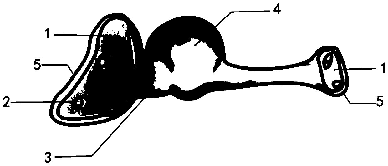
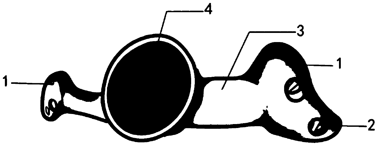
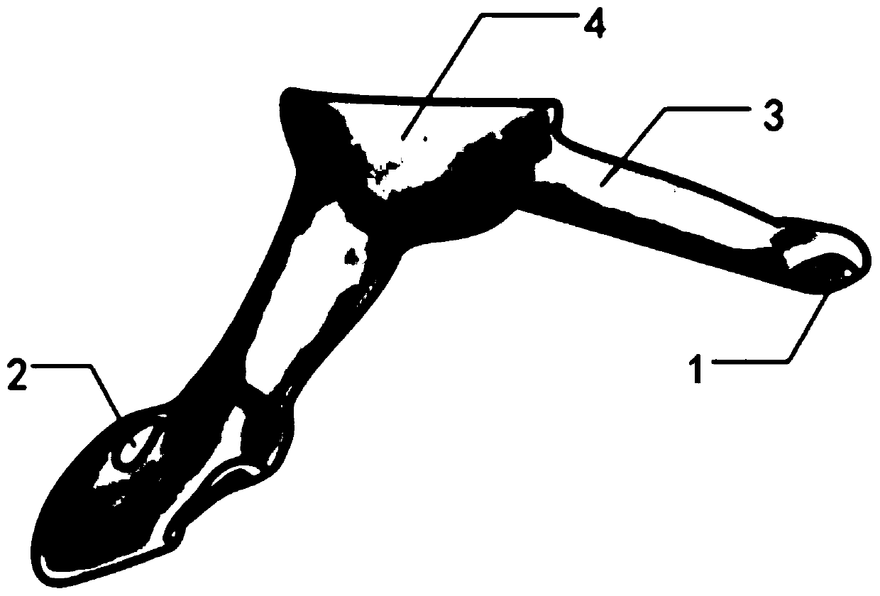
[0029] The present invention will be described in further detail below in conjunction with the accompanying drawings and embodiments.

[0030] see figure 1 , figure 2 , image 3 and Figure 4 The 3D printed hemipelvic prosthesis of the present invention is based on preoperative CT and MRI three-dimensional image fusion digital model for preoperative planning, to determine the scope of surgical resection, to obtain parameters such as pelvic osteotomy plane and acetabular position, and design according to these parameters Personalized prostheses for specific patients are produced, and after the design is completed, computer simulation is carried out to simulate the installation, and metal 3D printing equipment is used to prepare the actual prosthesis. During the operation, the 3D printed guide plate or computer-aided navigation system is used to complete the precise resection, and the osteotomy plane can fit perfectly with the contact surface of the prosthesis.

[0031] The...


PUM


Abstract

The invention discloses a 3D printing semi-pelvic prosthesis. The 3D printing semi-pelvic prosthesis comprises a prosthesis contact surface matched with the sacrum, a prosthesis contact surface matched with the contralateral pubis, a semi-circular acetabular cup structure and a cylindrical structure connecting the prosthesis contact surfaces and the semi-circular acetabular cup structure. The prosthesis contact surfaces at two ends are designed with porous structures, edge wrapping structures outside and two screw holes in the middle, and the cylindrical structures are designed by finite element mechanical analysis and topological structure optimization. The prosthesis contact surfaces at two ends, designed through early collected imaging data, can perfectly fit the sacrum and the contralateral pubis of a patient, respectively; the prosthesis and the contacted bone can be fixed through screws, the position of the center of the semi-circular acetabular cup structure after reconstructionis consistent with that of the original acetabulum center, so that better hip joint function and early stability can be achieved, and the porous structures of the prosthesis contact surfaces are beneficial to later bone ingrowth, so as to achieve long-term stability.

Description

technical field [0001] The invention relates to the manufacture of medical internal fixation implants, in particular to a 3D printed half pelvic prosthesis. Background technique [0002] The pelvis is the starting and ending point of a large number of muscles. There are abundant communication vessels and lack of natural tissue barriers. Malignant tumors that occur in bone tissue can easily penetrate bone and enter soft tissue, and tumors from soft tissue can also easily invade bone. Pelvic tumors are divided into primary tumors and metastatic tumors. The former is mainly seen in chondrosarcoma, osteosarcoma, and Ewing sarcoma; metastatic tumors are commonly seen in bone metastases from malignant tumors such as breast cancer, thyroid cancer, kidney cancer, prostate cancer, and lung cancer. Due to the complex anatomical structure and deep location of the pelvis, pelvic tumors often have an insidious onset. It often takes a long time for patients to go from the first symptoms ...

Claims


Application Information

Patent Timeline
IPC IPC(8): A61F2/30A61F2/32A61F2/34A61F2/44
CPCA61F2/30734A61F2/30771A61F2/30942A61F2/32A61F2/34A61F2/44A61F2002/30329A61F2002/30433A61F2002/30772A61F2002/30985
Inventor 范宏斌朱东泽付军王玲张煜珅孙立国刘鑫成党竞医
Owner FOURTH MILITARY MEDICAL UNIVERSITY
Why Eureka
Eureka Blog
Learn More