Quantitative liquid biopsy diagnostic system and methods

A buffer solution and blood sample technology, applied in the direction of measuring devices, material inspection products, fluorescence/phosphorescence, etc., can solve the problems of increasing method errors and tediousness

Inactive Publication Date: 2020-04-17
QCDX LLC
View PDF4 Cites 1 Cited by
  • Summary
  • Abstract
  • Description
  • Claims
  • Application Information

AI Technical Summary

Problems solved by technology

[0010] Therefore, it can be seen that the existing technology is limited by traditional solid tissue biopsy methods or CTC analysis systems that require cumbersome enrichment steps (which may increase the error of the method)

Method used


Image

  • Quantitative liquid biopsy diagnostic system and methods
  • Quantitative liquid biopsy diagnostic system and methods
  • Quantitative liquid biopsy diagnostic system and methods

Examples


Embodiment Construction

[0020] The present invention relates to a method for characterizing and quantifying target cells in a blood sample comprising the following steps:

[0021] (a) obtaining a blood sample from a subject,

[0022] (b) prepare the sample by one or more of the following steps (i) to (vi), including

[0023] (i) Centrifugation to separate the cell layer from the serum layer

[0024] (ii) removing one of the cell layers,

[0025] (iii) suspending the layer removed from step (b)(ii) in buffer,

[0026] (iv) purifying the suspension layer of (b)(iii),

[0027] (v) fixing the purification layer of (b)(iv), and

[0028] (vi) immunostaining and / or fluorescent in situ hybridization (FISH) staining of the fixed layer of (b)(v),

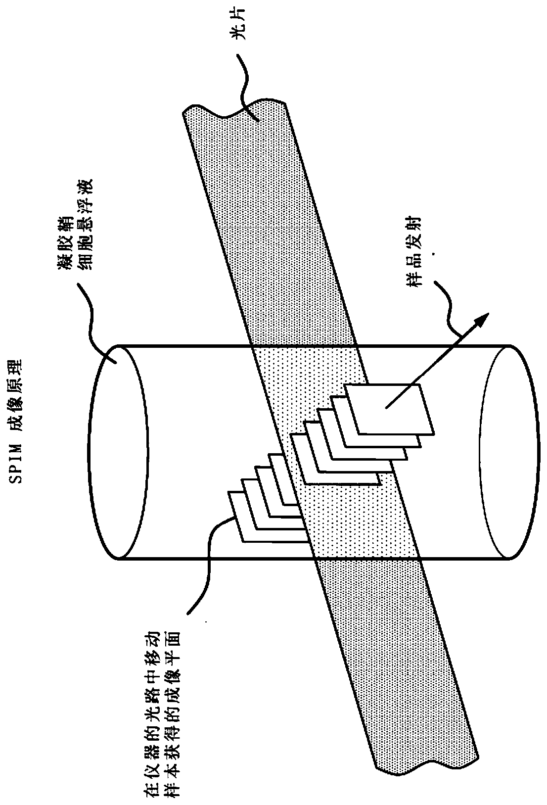
[0029] (c) placing the prepared sample from (b) under a selective planar image microscope and scanning the sample in multiple cross-sections by a light (eg laser) sheet source to obtain successive cross-sectional images,

[0030] (d) collect a sufficient numbe...


PUM

PropertyMeasurementUnit
diameteraaaaaaaaaa

Abstract

The present invention provides a quantitative liquid biopsy diagnostic system and methods for performing diagnostic assays. The system offers a liquid biopsy method using circulating tumor cells (CTCs) or White Blood Cells (WBC) subpopulations for precision cancer diagnosis, early detection of disease evolution, and cancer patient management. The invention utilizes selective plane illumination microscopy (SPIM) to deliver high sensitivity and specificity for the detection and isolation of individual CTCs, superseding the efficacy of existing methodologies for early cancer detection. Isolated CTCs can be analyzed for their molecular fingerprint, which can lead to matching genetic abnormalities with specific drug treatments. The system allows ex vivo observation of live CTC or WBC response to treatment. This observation of live cells offers the oncologist a new potential for optimizing therapeutic protocols by testing a patient's own cells, and then administering treatment to the patientwith the expectation of improving efficacy and reducing toxicity to normal cells.

Description

technical field [0001] The present invention provides a simple blood sample based quantitative liquid biopsy diagnostic system and method for performing diagnostic analysis. The system provides a liquid biopsy method that detects and identifies subpopulations of blood cells, including rare cells, such as circulating tumor cells (CTCs), derived from blood cells of a patient, for precise diagnosis of cancer, early detection of disease evolution, and cancer patient manage. The present invention utilizes Selective Plane Illumination Microscopy (SPIM) to provide high sensitivity and specificity for the detection and isolation of individual CTCs, replacing the efficacy of existing early cancer detection methods. Isolated CTCs can be analyzed for their molecular fingerprints, which can lead to matching genetic abnormalities with specific drug treatments. This system allows ex vivo observation of the response of living tumor cells to therapy. This observation of living cells offers...

Claims


Application Information

Patent Timeline
Patent Type & Authority Applications(China)
IPC IPC(8): G01N21/64G01N33/50G02B3/06G02B21/00
CPCG01N15/1468G01N33/50G01N21/6458G02B21/367G01N2015/1006G01N15/1434G01N2015/1445G02B21/06G02B21/16G01N1/2813G01N33/487
Inventor T·P·塔法斯S·温弗里
Owner QCDX LLC
Why Eureka
Try Eureka