CT image lung vessel segmentation method and system based on deep learning

A CT imaging and deep learning technology, applied in the field of medical image processing, can solve problems such as difficulty, low efficiency, and complex medical imaging results, and achieve the effect of improving accuracy

Active Publication Date: 2020-05-01
PERCEPTION VISION MEDICAL TECH CO LTD
View PDF5 Cites 17 Cited by
  • Summary
  • Abstract
  • Description
  • Claims
  • Application Information

AI Technical Summary

Problems solved by technology

Due to the complexity and individual differences of body tissues and organs, coupled with the differences between different imaging devices, the medical imaging results are very complicated, resulting in the inaccurate segmentation results of traditional medical image segmentation methods.
[0003] In the field of pulmonary vascular segmentation in the field of medical image segmentation, pulmonary vascular imaging has low contrast, complex structures of small blood vessels and trachea, and more noise in lung images, making accu

Method used


Image

  • CT image lung vessel segmentation method and system based on deep learning
  • CT image lung vessel segmentation method and system based on deep learning
  • CT image lung vessel segmentation method and system based on deep learning

Examples


Embodiment Construction

[0037] In order to make the purpose, technical solutions and advantages of the embodiments of the present invention clearer, the technical solutions in the embodiments of the present invention will be clearly and completely described below in conjunction with the drawings in the embodiments of the present invention. Obviously, the described embodiments It is a part of embodiments of the present invention, but not all embodiments. Based on the embodiments of the present invention, all other embodiments obtained by persons of ordinary skill in the art without creative efforts fall within the protection scope of the present invention.

[0038] Unless expressly stated otherwise, throughout the specification and claims, the term "comprise" or variations thereof such as "includes" or "includes" and the like will be understood to include the stated elements or constituents, and not Other elements or other components are not excluded.

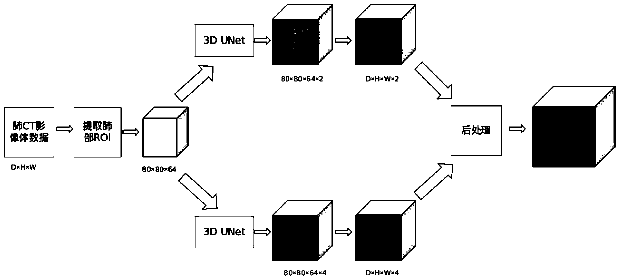
[0039] figure 1 A flow chart of a deep learnin...


PUM


Abstract

The embodiment of the invention provides a CT image pulmonary vessel segmentation method and system based on deep learning, a used deep learning network model is 3D UNet. The method comprises the following steps: S1, preprocessing; S2, performing three-dimensional sampling; S3, blood vessel segmentation; S4, distinguishing arteries and veins of the blood vessels; and S5, post-processing: combiningthe arteriovenous probability graph obtained by the arteriovenous distinguishing of the blood vessels and the blood vessel preliminary segmentation result obtained by the blood vessel segmentation module. According to the CT image pulmonary vessel segmentation method and system based on deep learning, intrapulmonary small vessels and extrapulmonary large vessels can be segmented, arteries and veins can be completely and automatically distinguished, and the segmentation accuracy is improved.

Description

technical field [0001] The present invention relates to the technical field of medical image processing, in particular to a method and system for segmenting pulmonary blood vessels in CT images based on deep learning. Background technique [0002] Lung cancer is the most threatening tumor to human life and health. Early detection is crucial to the survival and recovery of patients. Clinically, pulmonary nodule detection is the first step in lung cancer screening. Through the detection and segmentation of lung trachea and blood vessels in CT images, it is of great significance to the early screening and evaluation of lung cancer. Due to the complexity and individual differences of body tissues and organs, coupled with the differences between different imaging devices, the results of medical imaging are very complicated, resulting in the problem of inaccurate segmentation results of traditional medical image segmentation methods. [0003] In the field of pulmonary vascular s...

Claims


Application Information

Patent Timeline
IPC IPC(8): G06T7/11
CPCG06T2207/10081G06T2207/20081G06T2207/20084G06T2207/30061G06T2207/30101G06T2207/30204G06T7/11
Inventor 余明亮魏军
Owner PERCEPTION VISION MEDICAL TECH CO LTD
Why Eureka
Try Eureka