Isolation and application of a Salmonella phage rdp-sa-17118
A technology of RDP-SA-17118 and Salmonella, applied in the field of microorganisms, can solve the problems of drug failure and lack of antibiotics, and achieve the effect of good acid-base tolerance
- Summary
- Abstract
- Description
- Claims
- Application Information
AI Technical Summary
Problems solved by technology
Method used
Image
Examples
Embodiment 1
[0037] Example 1 Isolation and Identification of Pathogenic Salmonella Avian S6
[0038] Sampling from the diseased farm, aseptically take the liver of diseased poultry, streak it on the selective medium (SS agar), culture it at 37°C for 18-24 hours, and form a round, flat, neat edge and smooth surface on the medium Wet red colonies, pick typical colonies and continue to streak and purify 3 times, then pick a single colony and inoculate in 5mL LB broth, shake and culture at 200rpm at 37°C for 8h to obtain a uniform turbid bacterial suspension. Through 16sRNA molecular identification and serotype identification, it was determined to be pathogenic Salmonella, named S6, and stored in a -80°C refrigerator.
Embodiment 2
[0039] Example 2 Isolation and identification of phage RDP-SA-17118:
[0040](1) Manure treatment: Weigh 5 g of chicken manure and add it to 10 mL of sterile water to soak overnight, then centrifuge the overnight leachate at 10,000 rpm for 5 minutes, take the supernatant and pass it through a 0.22 μm filter, and use the filtrate for later use;
[0041] (2) Preparation of mixed bacterial suspension: Take 0.2mL of bacterial suspension and 0.1mL of filtrate and add it to 5mL of LB broth, culture at 37°C, shake at 200rpm overnight, then centrifuge at 10,000rpm for 5min, take the supernatant and pass it through a 0.22μm filter. Reserve the filtrate;
[0042] (3) Separation of bacteriophages: Separation of phages was carried out using the double-plate method. After mixing 0.1 mL of the filtrate of the mixed bacterial suspension and 0.2 mL of the host Salmonella suspension, they were bathed in water at 37°C for 10 minutes, and then double-plates were spread and placed at 37°C. After...
Embodiment 3
[0045] Electron microscope observation of embodiment 3 phage
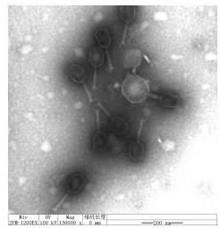
[0046] Take 20 μL of the liquid containing crude phage particles and drop them on the copper grid, let it settle naturally for 15 minutes, and absorb the excess liquid from the side with filter paper, add a drop of 2% phosphotungstic acid (PTA) on the copper grid to stain the phage for 10 minutes, and then use the filter paper to remove the Aspirate the staining solution from the side, and observe the phage morphology with an electron microscope after the sample is dry, as shown in figure 1 shown.
[0047] The bacteriophage RDP-SA-17118 has a polyhedral three-dimensional symmetrical head wrapped around nucleic acid, with a diameter of about 70nm, a tail about 120nm in length, a tail sheath, and a neck connecting the head and tail. According to the Ninth Report of the International Virus Taxonomy Organization Virus Classification, the bacteriophage is classified as Myoviridae of the order Cauviridae.
[0048] Whol...
PUM
| Property | Measurement | Unit |
|---|---|---|
| diameter | aaaaa | aaaaa |
Abstract
Description
Claims
Application Information
 - R&D
- Intellectual Property
- Life Sciences
- Materials
- Tech Scout
- Unparalleled Data Quality
- Higher Quality Content
- 60% Fewer Hallucinations
Browse by: Latest US Patents, China's latest patents, Technical Efficacy Thesaurus, Application Domain, Technology Topic, Popular Technical Reports.
© 2025 PatSnap. All rights reserved.Legal|Privacy policy|Modern Slavery Act Transparency Statement|Sitemap|About US| Contact US: help@patsnap.com


