

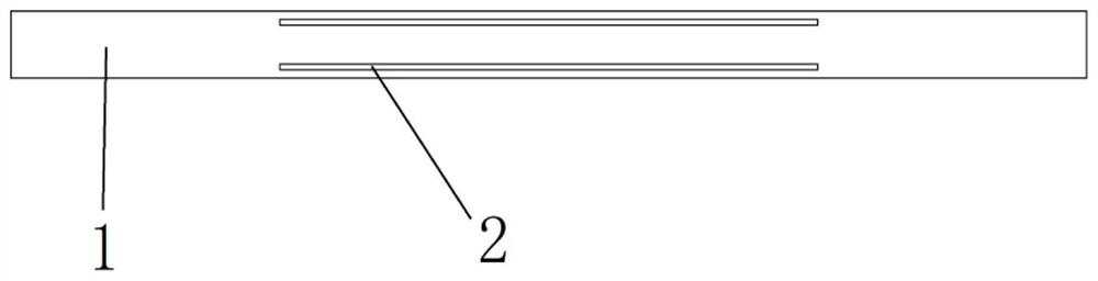
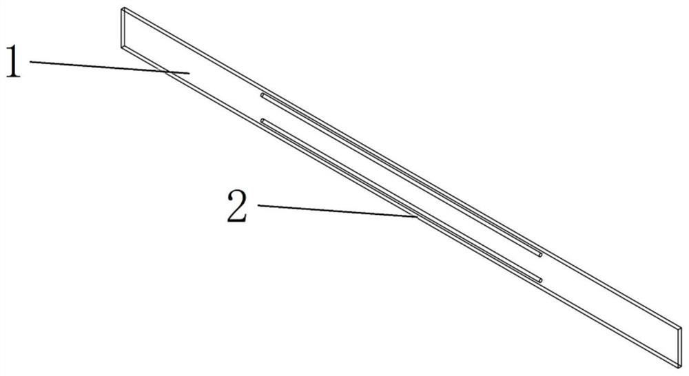
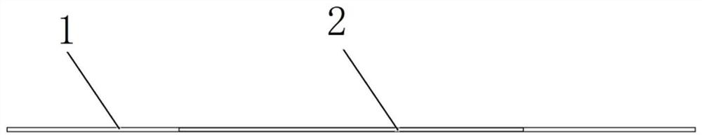
Posterior scleral reinforcement structures visualized by MRI
A technique of posterior sclera reinforcement and nuclear magnetic resonance, applied in the direction of eye implants, etc., can solve the problems that the surgical effect is difficult to guarantee and maintain, and does not consider the evaluation of the surgical effect after postoperative examination, and achieves good deformation resistance, easy operation, and protection eyeball effect
- Summary
- Abstract
- Description
- Claims
- Application Information
AI Technical Summary
Problems solved by technology
Method used
Image

Examples
Embodiment Construction
[0038] In order to make the purpose, technical solutions and advantages of the embodiments of the present invention more clear, the embodiments of the present invention will be further described in detail below in conjunction with the embodiments and the accompanying drawings. Here, the exemplary embodiments and descriptions of the present invention are used to explain the present invention, but not to limit the present invention.
[0039] In describing the present invention, it is to be understood that the terms "comprises / comprising", "consisting of" or any other variation thereof are intended to cover a non-exclusive inclusion such that a product, device, A process or method includes not only those elements, but may also include other elements not expressly listed, or elements inherent in such a product, apparatus, process, or method, as required. Without further limitations, an element defined by the phrase "comprising / comprising...", "consisting of... does not exclude the...
PUM
 Abstract
Description
Claims
Application Information