
Method for simultaneously detecting central demyelinating autoantibodies AQP4, MOG and MBP

An autoantibody, demyelination technology, applied in chemical instruments and methods, botanical equipment and methods, biochemical equipment and methods, etc., can solve the problems of unstable fluorescence efficiency, high cost, reduced detection sensitivity and specificity, etc. , to achieve the effect of improving labor capacity and quality of life, large social and economic benefits, and saving manpower and time costs

Pending Publication Date: 2020-06-12
天津天海新域生物科技有限公司 +1
View PDF11 Cites 2 Cited by
  • Summary
  • Abstract
  • Description
  • Claims
  • Application Information

AI Technical Summary

Problems solved by technology

It takes a long time, consumes a lot of manpower and material resources, and the fluorescence efficiency is unstable. A more stable transfection method is needed to obtain more sensitive and stable results.
The detection of MOG and MBP autoantibodies mostly uses nerve tissue sections with high expression of MBP antigen for staining and detection. There are many problems in this method, such as the difficulty in controlling the location of sectioned tissues, resulting in large differences between batches; most of the tissue sections are of animal origin (such as Monkey nerve tissue slices used by European companies), there are species differences in antigens, which reduce the sensitivity and specificity of detection; there are other antigens except MBP in the tissue, and the positive result of the test may not be positive for MBP antibody
2. Western or Elisa method detection, although the two technologies are relatively mature, but because it is difficult to maintain the spatial conformation of the MBP antigen, its specificity is not high, and the cost is expensive, which greatly limits the promotion of these detection methods

Method used


Image

  • Method for simultaneously detecting central demyelinating autoantibodies AQP4, MOG and MBP
  • Method for simultaneously detecting central demyelinating autoantibodies AQP4, MOG and MBP
  • Method for simultaneously detecting central demyelinating autoantibodies AQP4, MOG and MBP

Examples


Embodiment 1

[0027] Example 1, Construction of Stable HEK293T Cells Transferred with AQP4, MOG, and MBP Genes

[0028] 1. Carrier construction:

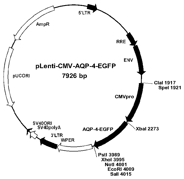
[0029] (1) Construction of an overexpression vector that fuses and expresses the human AQP-4 gene and the fluorescent marker gene EGFP

[0030] Using the pEGFP-N1 vector as a template, the primers EGFPF and EGFPR were used to amplify the EGFP sequence. The primers used were as follows: EGFPF: 5'-CTA GGATCC ATGGTGAGCAAGGGCGAGGAG-3' and EGFPR: 5'-CAGGTCGACGAATTCGCGGCCGCCTCGAGCTGCAGTTACTTGTACAGCTCGTCCATGCCG-3', and the amplified fragment was recovered by gel cutting Afterwards, it was connected to the T vector, and after the correct sequence was sequenced, it was double-digested with BamHI and SalI and connected to the pCDH-CMV-MCS-EF1-CopGFP carrier fragment after double-digestion with the same enzyme, and replaced with EGFP EF1-copGFP sequence in the original vector to obtain the plasmid vector pCDH-CMV-MCS-EGFP.

[0031] Using human optic nerve...

Embodiment 2

[0059] Embodiment 2, cell immunofluorescent staining assay serum AQP4, MOG, MBP antibody

[0060] 1) Cell inoculation: Mix cell attachment reagent (purchased from Beijing Xigong Biotechnology Co., Ltd., product number: 1027) with PBS at a volume ratio of 1:10, add to a 6-well plate containing a cover slip, 37°C, 5 %CO 2 Incubate for 1 hour. Then add stable HEK293T cell lines transfected with AQP4, MOG, and MBP genes respectively (ie, HEK293T cell lines containing AQP-4 genes, HEK293T cell lines containing MBP genes, and HEK293T cell lines containing MOG genes), and the cell inoculation amount is 1×10 6 / ml, 37°C, 5% CO 2 Cultivate to 80% confluence for subsequent detection. Under the same conditions, HEK293T cells transfected with empty vector were used as control.

[0061] 2) Cell immunofluorescence: cut the cover glass with a diamond engraving pen to a small square with a side length of 5mm, with the cell side facing up, paste it on the glass slide with shadowless glue,...

Embodiment 3

[0063] Embodiment 3, specific detection

[0064] In view of the low positive rate of MOG antibody detection, in order to further promote the clinical application of the method of the present invention, we selected 120 cases of serum from patients with neurological diseases in multiple centers and sent them to third-party companies in the EU for testing. detection. By comparing with the EU detection method, the MOG positive antibody detection rate of the method of the present invention was analyzed.

[0065] Choose 120 routine patients with nervous system disease serum (containing 30 cases of demyelinating patients), with the detection rate of Oumeng detection method (Oumeng Medical Diagnosis (China) Co., Ltd.) and the detection rate of the present invention's method (embodiment 2 method) The output rate is compared. The results showed that among the 30 demyelinating patients, Oumeng detected 4 cases of MOG antibody-positive sera, and this detection method detected 6 cases of...


PUM


Abstract

The invention discloses a method for simultaneously detecting central demyelinating autoantibodies AQP4, MOG and MBP. The method comprises the following steps of respectively introducing AQP4, MOG andMBP genes into HEK293T cells, respectively incubating the AQP4, MOG and MBP genes together with a sample to be detected, detecting by adopting an immunofluorescence method and taking the HEK293T cells transfected with an empty carrier as a contrast, and determining that the sample to be detected is positive if a fluorescence signal is stronger than the contrast; and determining that the sample tobe detected is negative if the fluorescence signal is equal to or smaller than the control signal. The invention provides the method for simultaneously detecting central demyelinating autoantibodiesAQP4, MOG and MBP in a same system, a fluorescence immunocyte staining technology with higher sensitivity and specificity is used for clinically and qualitatively detecting AQP4, MOG and MBP antibodies, so that the detection result is more stable, and the labor and time costs are greatly saved.

Description

[0001] (1) Technical field [0002] The invention relates to a detection of central demyelinating autoantibodies, in particular to a method for simultaneously detecting central demyelinating autoantibodies AQP4, MOG and MBP. [0003] (2) Background technology [0004] Demyelinating disease of the central nervous system is an autoimmune disease mainly characterized by multifocal and inflammatory demyelination of the central nervous system, and its clinical features mainly include recurrent attacks, multiple remissions and relapses. The more common clinical demyelinating diseases of the central nervous system include multiple sclerosis (Multiple Sclerosis, MS), neuromyelitis optica (neuromyelitis optica, NMO), acute disseminated encephalomyelitis and so on. In recent years, the incidence of central nervous system demyelinating diseases has been increasing year by year, which has a serious impact on the quality of life of patients and poses a serious threat to patients' physical a...

Claims


Application Information

Patent Timeline
IPC IPC(8): G01N33/543G01N33/533C12N15/867C12N15/13C12N5/10
CPCG01N33/543G01N33/533C07K14/47C07K16/00C12N15/86C12N2740/15043
Inventor 施福东李敏淑金薇娜方朝君金立方么阳贾冬梅
Owner 天津天海新域生物科技有限公司
Learn More