
Exosome derived from human umbilical cord mesenchymal stem cells, and identification method and application of exosome

A technology of stem cells and identification methods, applied in the field of human umbilical cord mesenchymal stem cell-derived exosomes, identification and application, can solve the problems of tumorigenic risk, ethical issues, immune rejection, etc., to improve the disease, improve the heart function, the effect of inhibiting apoptosis

Active Publication Date: 2020-07-31
THE SECOND HOSPITAL AFFILIATED TO WENZHOU MEDICAL COLLEGE
View PDF7 Cites 5 Cited by
  • Summary
  • Abstract
  • Description
  • Claims
  • Application Information

AI Technical Summary

Problems solved by technology

However, it has been found that the use of stem cell transplantation treatment has problems such as immune rejection, ethical issues, and tumorigenic risks.

Method used


Image

  • Exosome derived from human umbilical cord mesenchymal stem cells, and identification method and application of exosome
  • Exosome derived from human umbilical cord mesenchymal stem cells, and identification method and application of exosome
  • Exosome derived from human umbilical cord mesenchymal stem cells, and identification method and application of exosome

Examples


Embodiment 1

[0024] Example 1: Obtaining of human umbilical cord mesenchymal stem cell exosomes (hucMSC-exosomes)

[0025] Step 1: Isolation and culture of human umbilical cord mesenchymal stem cells (hucMSCs):

[0026] Human umbilical cord mesenchymal stem cells (hucMSCs) were obtained by collagenase digestion. Briefly, under aseptic conditions, the umbilical cord tissue was dissected and the umbilical arteries and veins were removed. The umbilical cord tissue was washed twice with PBS containing 1% gentamicin to remove blood clots and red blood cells, and cut into 1mm 3 size organization block. Then digest with 0.1% type II collagenase on a shaker at 37°C for 6 hours, filter with a 100-mesh screen, centrifuge the filtrate at 1000 rpm for 10 minutes, resuspend the pellet in PBS and wash twice. Use DMEM / F12 + 10% FBS (fetal bovine serum) + 1% P / S (penicillin / streptomycin) complete medium to gently blow the cell pellet, mix well, and transfer to 25cm 2 in a petri dish at 37°C, 5% CO 2 ...

Embodiment 2

[0029] Example 2: Identification of human umbilical cord mesenchymal stem cell exosomes (hucMSC-exosomes)

[0030] Human umbilical cord mesenchymal stem cells (hucMSCs) obtained in Step 1 of Example 1 were identified by flow cytometry: the third generation hucMSCs (P3) were digested with trypsin containing 0.25% EDTA, and centrifuged at 1500 rpm for 5 minutes to collect hucMSCs. Wash once with PBS, then fix with 500 microliters of 4% paraformaldehyde overnight at 4°C, discard the fixative, resuspend with 2 milliliters of PBS, and centrifuge to discard the supernatant. Block with 2 ml of PBS containing 5% FBS for 10 minutes at room temperature, centrifuge to discard the supernatant, and set an isotype control group at the same time. Add FITC or PE-labeled fluorescent antibodies FITC-CD29, FITC-CD44, FITC-CD59, FITC-CD90, PE-CD105, PE-CD166, and FITC-CD34 working solution 100 μl at a ratio of 1:50 (5 μl antibody / 100 microliters of cell suspension), incubate at room temperature i...

Embodiment 3

[0032] Example 3: Verification of the effect of human umbilical cord mesenchymal stem cells (hucMSCs) in the treatment of viral myocarditis (VMC)

[0033] Virus

[0034] Coxsackie B3 virus (CVB3, Nancy strain, USA) was maintained and amplified using Hep2 cells. Virus titers were determined by the 50% tissue culture infectious dose (TCID50) method.


PUM

PropertyMeasurementUnit
diameteraaaaaaaaaa

Abstract

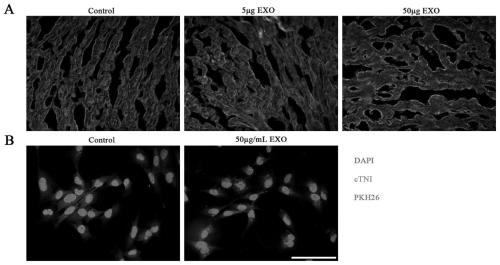
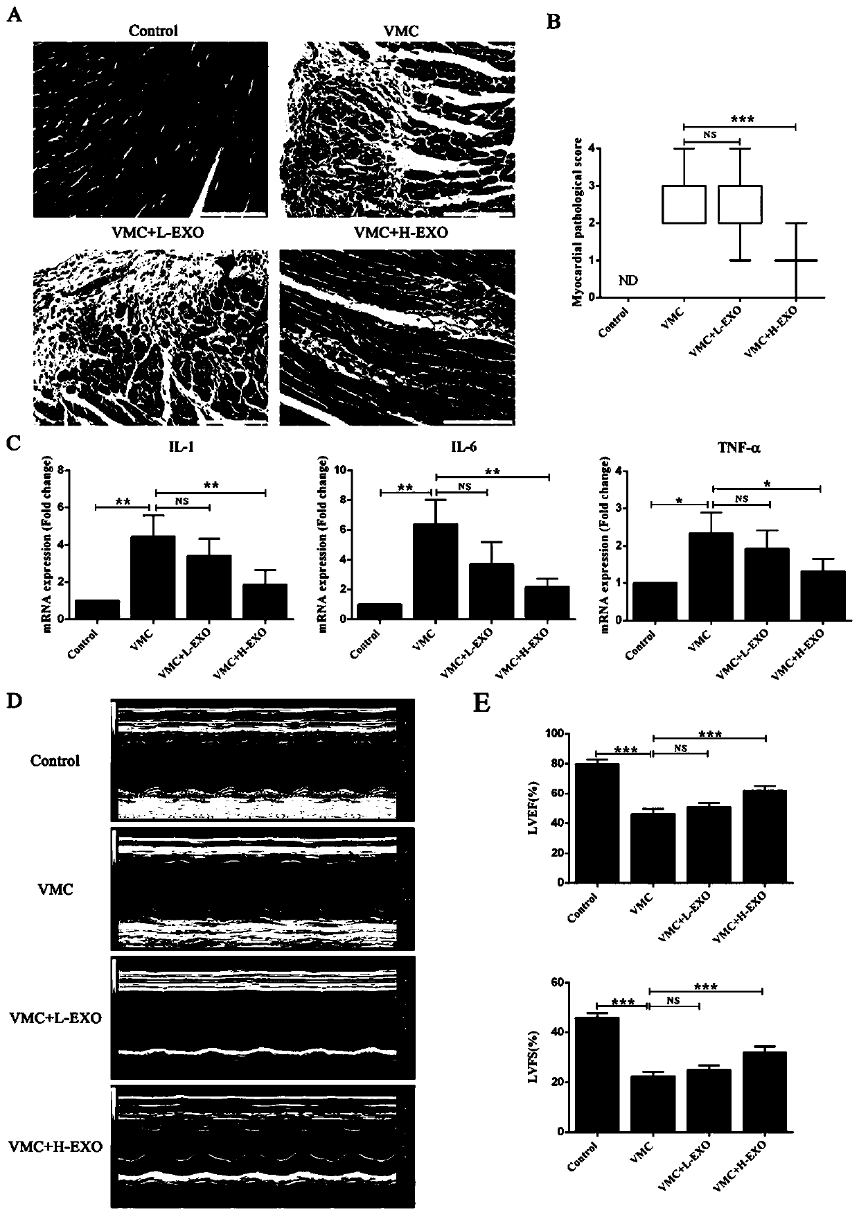
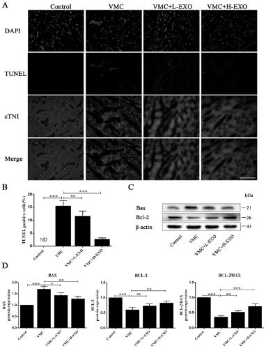
The invention relates to an exosome derived from human umbilical cord mesenchymal stem cells, and an identification method and application of the exosome. The present invention finds that the exosomesecreted by human umbilical cord mesenchymal stem cells can be ingested by myocardial cells, and can effectively improve the cardiac function of coxsackie B3 (CVB3) induced viral myocarditis mice (VMC), inhibit apoptosis of cardiac cells of the myocarditis mice, and inhibited inhibit apoptosis of human cardiac muscle cells (HCMs) cultured in vitro under induction of CVB3. According to the application disclosed by the invention, the exosome derived from human umbilical cord mesenchymal stem cells can act on a VMC mouse model, so that the effect of improving diseases is achieved to a certain extent, and a new method is provided for treating viral myocarditis by adopting the stem cell-derived exosome in the future.

Description

technical field [0001] The invention relates to an exosome derived from human umbilical cord mesenchymal stem cells, an identification method and an application. Background technique [0002] Viral myocarditis (VMC) is an important cause of heart failure, arrhythmia, and sudden death in young people. This disease is closely related to the immune damage and apoptosis of the body cells caused by virus infection. In the past few decades, patients with viral myocarditis have been treated with conventional treatment, immunomodulatory treatment, immunosuppressive treatment, etc., which can partially bring about clinical recovery, but patients who cannot fully recover may develop dilated cardiomyopathy. Therefore, it is becoming more and more necessary to explore new effective treatments. [0003] Stem cells are primitive cells with the potential of self-replication and multi-directional differentiation. They are the origin cells of the body and the progenitor cells that form vari...

Claims


Application Information

Patent Timeline
Patent Type & Authority Applications(China)
IPC IPC(8): C12N5/0775G01N33/68A61K35/51A61P9/00A61P31/12
CPCA61K35/51A61P9/00A61P31/12C12N5/0665C12N2509/00C12N2509/10G01N33/6872
Inventor 郭晓令顾孝红褚茂平
Owner THE SECOND HOSPITAL AFFILIATED TO WENZHOU MEDICAL COLLEGE
Learn More