Dynamic enhanced magnetic resonance imaging processing method and system, storage medium and terminal

A magnetic resonance imaging and dynamic enhancement technology, which is applied in image data processing, image enhancement, neural learning methods, etc., can solve the problems of less deep learning research, inability to apply breast cancer diagnosis, difficulties, etc.

Active Publication Date: 2020-07-31
NORTHWEST UNIV
View PDF3 Cites 9 Cited by
  • Summary
  • Abstract
  • Description
  • Claims
  • Application Information

AI Technical Summary

Problems solved by technology

[0004] (1) The process of traditional machine learning methods is complex and different, and cannot be extended and applied in this field
[0005] (2) Dynamic enhanced magnetic resonance imaging technology contains multiple images with time series, and the existing methods do not make full use of its multi-phase image information, resulting in poor diagnostic results
[0006] (3) There are few studies on deep learning in this field, and the proposed network is relatively simple, which cannot be applied to the diagnosis of breast cancer with various information
[0008] (1) For the diagnosis of breast cancer based on traditional machine learning, due to the complex process, effective feature extraction, feature selection methods and the adoption of suitable classifiers are relatively difficult
[0009] (2) The dynamic enhanced magnetic resonance imaging technology has multi-phase images, and there is also a timing relationship between the images. How to mine the timing information of multi-phase images in the system is very difficult
[0010] (3) The research on the application of deep learning in breast cancer is still in its infancy, and how to design the network so that it can be used for various information of breast cancer diagnosis is more difficult

Method used


Image

  • Dynamic enhanced magnetic resonance imaging processing method and system, storage medium and terminal
  • Dynamic enhanced magnetic resonance imaging processing method and system, storage medium and terminal
  • Dynamic enhanced magnetic resonance imaging processing method and system, storage medium and terminal

Examples


Embodiment Construction

[0071] In order to make the object, technical solution and advantages of the present invention more clear, the present invention will be further described in detail below in conjunction with the examples. It should be understood that the specific embodiments described here are only used to explain the present invention, not to limit the present invention.

[0072] Aiming at the problems existing in the prior art, the present invention provides a dynamic enhanced magnetic resonance imaging processing method, system, storage medium, and terminal. The present invention will be described in detail below with reference to the accompanying drawings.

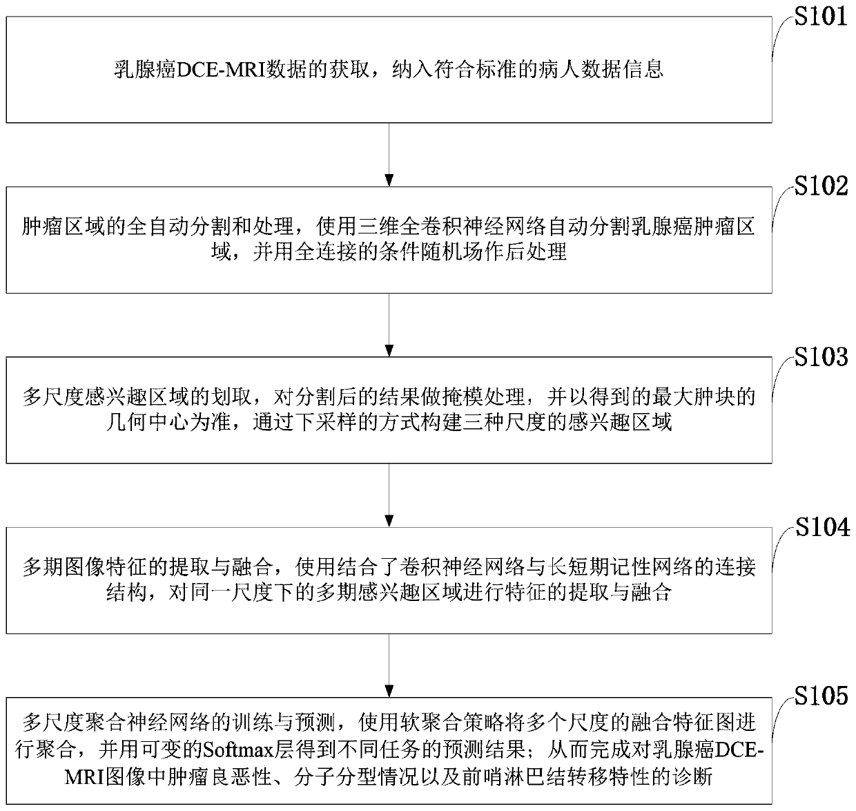
[0073] like figure 1 As shown, the dynamic enhanced magnetic resonance imaging processing method provided by the present invention comprises the following steps:

[0074] S101: Acquisition of breast cancer DCE-MRI data, including patient data information that meets the standards;

[0075] S102: Fully automatic segmentation and proces...


PUM


Abstract

The invention belongs to the technical field of medical image processing and computer-aided diagnosis, and discloses a dynamic enhanced magnetic resonance imaging processing method and system, a storage medium and a terminal, and the method comprises the steps: carrying out the full-automatic segmentation and processing of a primary tumor region in an obtained breast cancer DCE-MRI multi-stage image; carrying out mask processing on the segmented result, and taking the geometric center of the obtained maximum lump as a criterion to draw regions of interest of three scales; meanwhile, sending the multi-stage region-of-interest images under three scales into three connection structures combining a convolutional neural network and a long-term and short-term memory network for feature extraction and fusion; and sending the multi-stage fusion features obtained under the three scales into a soft aggregation model for aggregation of the overall features to obtain prediction results of different tasks. According to the method, the limitation of a shallow architecture is overcome, and the prediction precision is greatly improved; and perfect preoperative reference information can be visuallyprovided for operative treatment of breast cancer patients.

Description

technical field [0001] The invention belongs to the technical fields of medical image processing and computer-aided diagnosis, and in particular relates to a dynamic enhanced magnetic resonance imaging (DCE-MRI) processing method, system, storage medium and terminal. Background technique [0002] At present, breast cancer is the most common malignant tumor affecting women all over the world, and its high morbidity and mortality seriously threaten the health of women all over the world. If the relevant information of breast cancer tumors can be detected and diagnosed early, so as to formulate personalized treatment methods, the mortality rate of breast cancer patients can be greatly reduced and good results can be achieved. Preoperative determination of benign and malignant breast cancer tumors, molecular classification, and sentinel lymph node metastasis characteristics are all important factors for surgical treatment arrangements for patients. At present, radiologists usual...

Claims


Application Information

Patent Timeline
Patent Type & Authority Applications(China)
IPC IPC(8): G06T7/10G06T5/00G06N3/04G06N3/08
CPCG06T7/10G06T5/001G06N3/08G06T2207/10096G06T2207/20081G06T2207/20104G06T2207/20172G06T2207/20221G06T2207/30068G06T2207/30096G06N3/047G06N3/045
Inventor 侯榆青邓鹏飞赵凤军陈吉新董迪迪周昊文陈欣贺小伟
Owner NORTHWEST UNIV
Why Eureka
Try Eureka