Preparation method and application of immunomodulatory extracellular vesicles

An immunomodulatory, vesicle technology for medical applications

Active Publication Date: 2020-10-09
NANTONG UNIVERSITY
View PDF2 Cites 2 Cited by
  • Summary
  • Abstract
  • Description
  • Claims
  • Application Information

AI Technical Summary

Problems solved by technology

To sum up, the known tumor immunotherapy is mainly aimed at the established tumors and is related to the activation of T lymphocytes. Immunotherapy represented by PD-1/PD-L1 and CAR-T therapy

Method used


Image

  • Preparation method and application of immunomodulatory extracellular vesicles
  • Preparation method and application of immunomodulatory extracellular vesicles
  • Preparation method and application of immunomodulatory extracellular vesicles

Examples


preparation example Construction

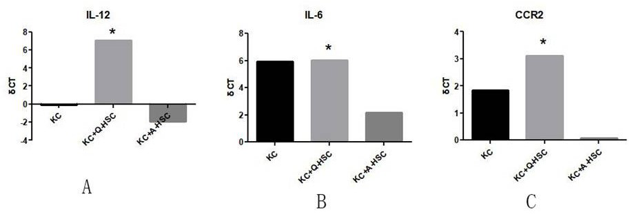
[0028] The present invention provides a preparation method of immunoregulatory small extracellular vesicles, which can activate macrophages and transform to M1 phenotype. The preparation method comprises the following steps:

[0029] (a) culturing the primary isolated and purified animal cells in a suitable culture system for a period of time, so as to accumulate an effective concentration of immunoregulatory small extracellular vesicles in the culture medium;

[0030] (b) collecting the culture medium of step (a);

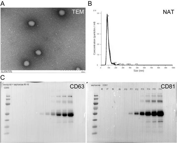
[0031] (c) Isolation of immunomodulatory small extracellular vesicles from the culture medium collected in step (b).

[0032] Wherein, the suitable culture system refers to: within 3 days after the primary separation and purification, the normal interface of the two-dimensional system is cultivated; and low-sugar DMEM culture medium) to simulate the physiological microenvironment in vivo, so that the physiological behavior of cells is closer to the actual physiol...

Embodiment 1

[0071] Example 1: Isolation of Cells from Healthy Tissue

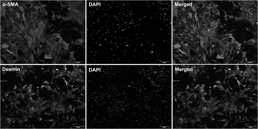
[0072] Isolation of hepatic stellate cells from healthy rat liver.

[0073] 1. Liver in situ perfusion, 0.075% type IV collagenase, digested at 37°C for 35 minutes;

[0074] 2. Separate the liver, shred it, and digest it with 0.015% type IV collagenase at 37°C for 15 minutes;

[0075] 3. Resuspend the D-Hanks, centrifuge the sample at 50g for 10min, and take the supernatant;

[0076] 4. Centrifuge at 500g for 10min to collect the precipitate;

[0077] 5. Centrifuge with a final concentration of 11-13% Nycodenz density gradient to collect cells in the middle layer;

[0078] 6. Cell counting and trypan blue staining;

[0079] 7. Plate the freshly isolated cells in a 5×10 5 Inoculate at 25cm per ml 2 In the culture flask, use Eagle's minimum basic low-glucose medium (containing 2% fetal bovine serum, 10mL / L Pen / Strep solution, 2mM Ala-Gln solution).

Embodiment 2

[0080] Example 2: Culture of isolated cells under simulated in vivo physiological conditions

[0081] For example, cells are grown by inoculating the medium at approximately 500,000 cells per milliliter.

[0082] The cells obtained in Example 1 are cultured on the ordinary two-dimensional interface until the third day, and the medium is collected; or incubated in a suitable simulated organ three-dimensional culture environment and a suitable medium. By using three-dimensional Matrigel and low-glucose culture conditions at 37°C, the physiological state in vivo is simulated. After culturing for a period of time sufficient to accumulate useful levels of immunomodulatory small extracellular vesicles, eg, up to about 6 days, the cultured cells and medium are harvested.


PUM


Abstract

The invention provides a preparation method and application of immunomodulatory extracellular vesicles, and the preparation method comprises the following steps: (a) culturing animal cells subjected to primary separation and purification in a culture system for a period of time so as to accumulate effective concentration of immunomodulatory extracellular vesicles in a culture medium; (b) collecting the medium of step (a); and (c) separating the immunomodulatory extracellular vesicles from the culture medium collected in the step (b). The extracellular vesicle provided by the invention has immunomodulatory activity, can activate macrophages and convert into M1 phenotype, and provides a theoretical basis for prevention or treatment of diseases caused or aggravated by immunosuppression.

Description

technical field [0001] The invention belongs to the field of medical technology, and in particular relates to a preparation method and application of immunoregulatory small extracellular vesicles. Background technique [0002] Immunity refers to a protective response of biological organisms to recognize and eliminate antigenic substances, including specific immunity and non-specific immunity. , chlamydia, parasites, etc.) and other harmful substances; immune surveillance, to discover and eliminate "non-self" components that appear in the body at any time; such as tumor cells and aging and apoptotic cells that occur due to gene mutations; immune self-stabilization, through self- Tolerance and immune regulation are two major mechanisms to achieve homeostasis within the immune system. In 1909, Paul Ehrlich proposed that the immune system can curb tumor occurrence, and abnormal immune function is the basic cause of tumor occurrence. In 1959, Frank Macfarlane Burnet and Lewis T...

Claims


Application Information

Patent Timeline
IPC IPC(8): C12N5/078A61K35/12A61K35/407A61K45/06A61P37/04
CPCC12N5/0634A61K35/12A61K35/407A61K45/06A61K9/0019A61K9/007A61P37/04C12N2513/00
Inventor 季菊玲孙玉风陆鹏程丽刘玮琪张安琪季煜华吕秀芳
Owner NANTONG UNIVERSITY
Why Eureka
Try Eureka