Molecular marker for early screening of breast cancer and application thereof
A molecular marker and breast cancer technology, applied in the field of medicine and biology, can solve the problems of lack of specific early molecular diagnosis methods and great harm, and achieve significant economic and social benefits, low misdiagnosis rate, and high sensitivity
- Summary
- Abstract
- Description
- Claims
- Application Information
AI Technical Summary
Problems solved by technology
Method used
Image


Examples
Embodiment 1
[0028] Example 1. Preparation and production of antibodies against phosphorylated 53BP1 protein
[0029] 1. Prepare antibodies against phosphorylated 53BP1 protein by immunizing animals:
[0030] First, design the polypeptides required to prepare antibodies based on the phosphorylation site of 53BP1; design and prepare the phosphorylation site (the 25th and 29th serines) of the protein kinase ATM (ataxia-telangiectasia mutated gene) phosphorylation site in the phosphorylated 53BP1 protein molecule The 53BP1 antigen polypeptide CIED(pS)QPE(pS)QVLEDD, wherein (pS) is serine with phosphate, was synthesized by Hangzhou Dangang Biological Company.
[0031] The phosphorylated 53BP1 antigen polypeptide was coupled to the carrier protein KLH (hemocyanin). Then the antigen solution and the antibody adjuvant (Freund's complete adjuvant or Freund's incomplete adjuvant, Sigma company product) were mixed at a volume ratio of 1:1, and subcutaneously injected into the back of New Zealand wh...
Embodiment 2
[0036] Embodiment 2, the assembly of molecular diagnostic kit and sample test
[0037] 1. Kit assembly
[0038] A specific immunohistochemical staining kit was assembled based on the specific antibody prepared in Example 1. In addition to the above-mentioned specific antibodies, the kit also has the following components:
[0039] Reagent A. Ready-to-use blocking solution, 1 bottle (3ml) of 10% non-immune goat serum
[0040] Reagent B. 1 bottle (3ml) of ready-to-use biotin-labeled goat anti-rabbit secondary antibody (Beyond Biotech, Cat. No. A0277)
[0041] Reagent C. 1 bottle of ready-to-use streptavidin peroxidase (3ml)
[0042] Reagent D. Concentrated Substrate Buffer (20X) 1 bottle (160ul)
[0043] Reagent E.DAB chromogen (20X) 1 bottle (160ul)
[0044] Reagent F. 0.6% H 2 o 2 (20X) 1 bottle (160ul)
[0045] 2. Clinical trials of kits
[0046] Purchase breast cancer tissue chips from Shanghai Zhuohao Pharmaceutical Technology Co., Ltd. (article number BRC2281, see at...
Embodiment 3
[0067] Embodiment 3, sample immunohistochemical staining result analysis
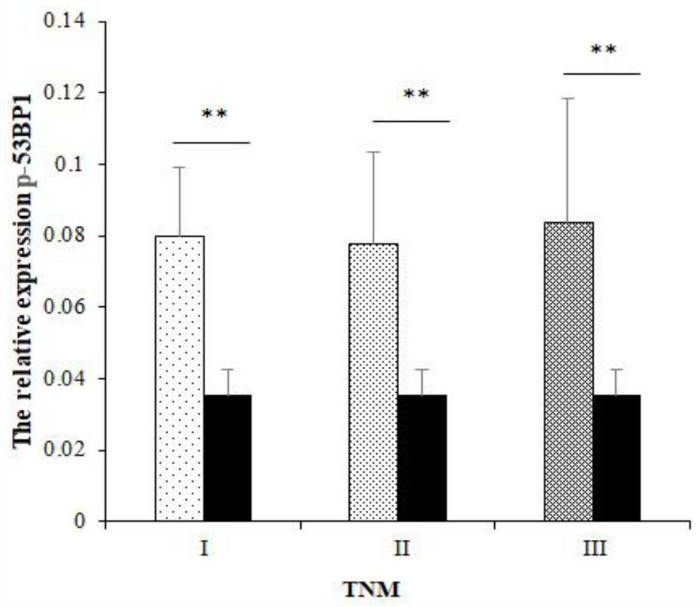
[0068] Image scope X64 software was used to collect immunohistochemical photos, Image pro plus software was used to analyze the results of immunohistochemical staining, and the IOD value of each picture was calculated. The data were statistically analyzed using SPSS 17.0 software, and the difference was considered statistically significant when P<0.01 was tested by an independent sample T test.
[0069] Through immunohistochemical staining technology, the above-mentioned specific phosphorylated 53BP1 antiserum was used to perform immunohistochemical staining on breast cancer tissue chips. A total of 228 cases were stained. The staining results are as follows: figure 1 As shown, the darker the color, the higher the concentration of the target antigen in the sample. Among them, A1, A2, A3, A4, A5, A6, A7, and A8 were 8 non-breast cancer tissue cases, and there were 220 breast cancer tissue cases. In the...
PUM
| Property | Measurement | Unit |
|---|---|---|
| molecular weight | aaaaa | aaaaa |
Abstract
Description
Claims
Application Information