Eureka AIR delivers breakthrough ideas for toughest innovation challenges, trusted by R&D personnel around the world.

Ultrasonic endoscopic image intelligent segmentation and quantification method and system based on deep learning

An endoscopic image and deep learning technology, applied in the field of biomedical image processing and deep learning, can solve problems such as difficult to obtain segmentation results, easy to detect false boundaries, image information loss, etc., to achieve easy promotion and application, compact structure, The effect of improving accuracy

Active Publication Date: 2020-10-16
SOUTH CHINA NORMAL UNIVERSITY
View PDF6 Cites 8 Cited by
  • Summary
  • Abstract
  • Description
  • Claims
  • Application Information

AI Technical Summary

Problems solved by technology

The grayscale threshold-based segmentation method is the most common image segmentation method that directly detects the region. One or more thresholds are used to segment the image into multiple target areas or backgrounds. In order to distinguish the target, each area needs to be marked later, but this The method is not suitable for images with small grayscale differences or large overlaps in the grayscale value range of each target area, and it is difficult to obtain accurate segmentation results; the basic idea of ​​​​the region growing method is to merge pixels with similar properties together, A region must first designate a seed point as the starting point of growth, then compare the pixels in the neighborhood around the seed point with the seed point, merge points with similar properties and continue to grow outward until no pixels that meet the conditions are included So far, this method requires manual interaction to obtain seed points, and a seed point must be implanted in each region that needs to be extracted. At the same time, the region growing method is very sensitive to noise, resulting in holes in the extracted regions or will be separated. The edge detection method is to solve the image segmentation problem by detecting the edge between different regions. The gray value of the pixel on the edge of the region often changes drastically. The edge detection operator is very sensitive to the edge information, and it is easy to detect false Boundaries, and are also very sensitive to pixels, usually filter the image before applying the detection operator, and filtering may lead to loss of image information

Method used


Image

  • Ultrasonic endoscopic image intelligent segmentation and quantification method and system based on deep learning
  • Ultrasonic endoscopic image intelligent segmentation and quantification method and system based on deep learning
  • Ultrasonic endoscopic image intelligent segmentation and quantification method and system based on deep learning

Examples


Embodiment Construction

[0045] In order to enable those skilled in the art to better understand the solutions of the present application, the technical solutions in the embodiments of the present application will be clearly and completely described below in conjunction with the drawings in the embodiments of the present application. Obviously, the described embodiments are only a part of the embodiments of the present application, rather than all the embodiments. Based on the embodiments in this application, all other embodiments obtained by those skilled in the art without creative work shall fall within the protection scope of this application.

[0046] The intelligent segmentation and quantification method of ultrasound endoscopic images based on deep learning of the present invention improves the accuracy of image segmentation based on the deep learning method. By using this method, only the collected ultrasound endoscopic images are subjected to simple preprocessing. It can be input into the neural...


PUM


Abstract

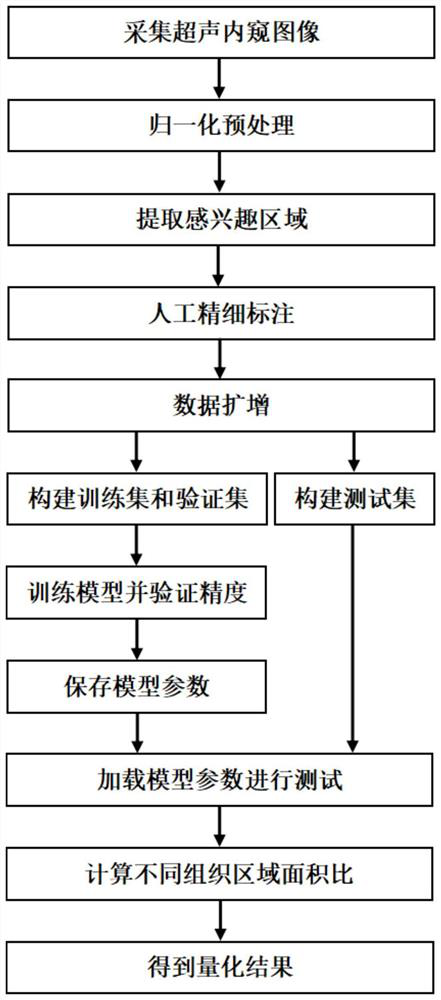
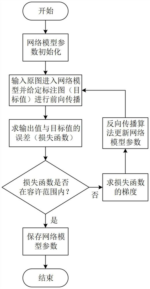
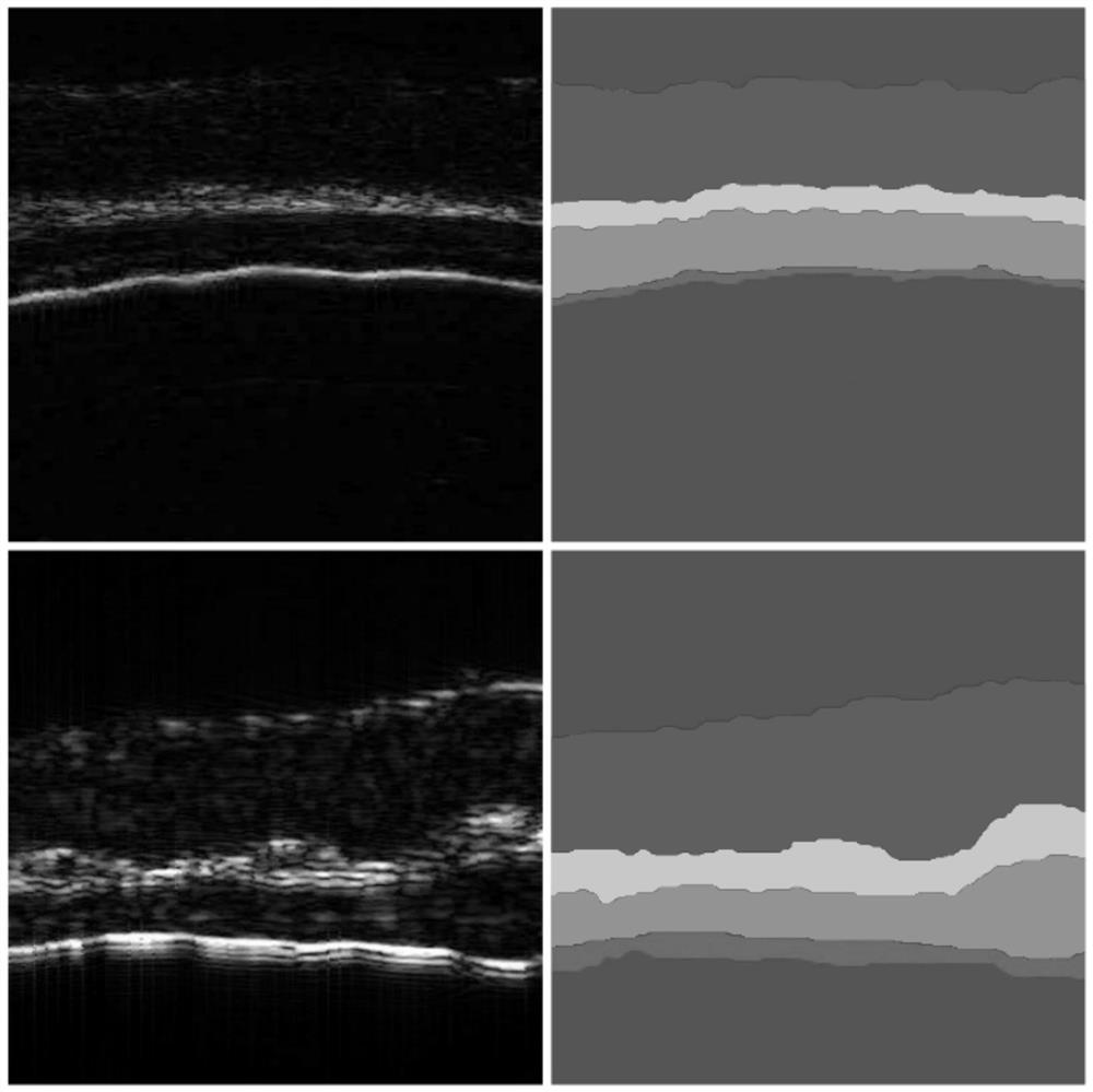
The invention discloses an ultrasonic endoscopic image intelligent segmentation and quantification method and system based on deep learning. The method comprises the following steps: carrying out image normalization preprocessing; extracting a region of interest; carrying out artificial fine labeling on different acoustic impedance layers; performing data amplification to obtain more images, and dividing the images and the corresponding manually labeled images into a training set, a verification set and a test set; constructing a full convolutional neural network model; sending the training set into a network model for training to obtain a segmentation model; verifying the segmentation precision of the trained model for the verification set; and calculating the relative area ratio of the acoustic impedance layers of different tissues after segmentation to obtain a quantification result. Based on the method, a fine acoustic impedance layered segmentation image and accurate quantizationparameters can be obtained, labor cost is reduced, and the method is expected to be applied to the fields of medical image analysis and the like.

Description

Technical field [0001] The invention belongs to the field of biomedical image processing and deep learning, and specifically relates to a method and system for intelligent segmentation and quantification of ultrasound endoscopic images based on deep learning. Background technique [0002] With the popularization and application of ultrasound endoscopes, high-resolution, large-scale, and high-depth ultrasound images play a very important role. Ultrasound endoscopic imaging is based on detecting the echo of the ultrasound signal in the tissue. It can image the tissue level and nearby organs, reflect the difference of tissue acoustic impedance, and can be used to check deep information. Ultrasound endoscopic image segmentation can identify and extract the boundaries of the acoustic impedance layer of different tissues, and then intuitively distinguish the differences and boundaries of each layer, and quantify the relative area ratio of each layer of tissue structure. [0003] Existin...

Claims


Application Information

Patent Timeline
IPC IPC(8): G06T7/13G06T7/11G06N3/08G06N3/04
CPCG06T7/13G06T7/11G06N3/08G06N3/045
Inventor 杨思华李陵熊科迪
Owner SOUTH CHINA NORMAL UNIVERSITY
Why Eureka
Eureka Blog
Learn More