Cellular image analysis method, cellular image analysis device, and learning model creation method

An image analysis and learning model technology, applied in the field of analysis and processing, can solve the problems of missing bad areas or foreign objects, poor efficiency of cell evaluation, and low judgment accuracy

Pending Publication Date: 2020-10-27
SHIMADZU SEISAKUSHO CO LTD
View PDF6 Cites 3 Cited by
  • Summary
  • Abstract
  • Description
  • Claims
  • Application Information

AI Technical Summary

Problems solved by technology

There are various feature quantities that can be used for such determination, and an appropriate feature quantity should be selected from among them in advance, but when the cell species or culture conditions to be observed are different, there is an optimum method for determining the state of the cell, etc. The case where the appropriate feature quantity changes
Therefore, in order to always make high-precision judgments, it is necessary to change the feature data used depending on the cell type to be observed, the culture conditions, etc., and the processing is very troublesome.
In addition, until then, when judging cells cultured under different conditions, it was necessary to study which characteristic quantity is suitable for the judgment, and there was also a problem that the efficiency of cell evaluation was low.
[0010] Moreover, in the above-mentioned existing cell evaluation methods, false images of feeder cells or background regions, or foreign objects such as impurities are often misjudged as target cells. Although the above-mentioned processing is cumbersome and the judgment accuracy is not high
In addition, since it is necessary to divide the image into small areas of multiple pixel units and make a judgment on each small area based on the limitation of the calculation time, it is possible to miss the small area that is very small compared to the size of the small area bad areas or foreign objects

Method used


Image

  • Cellular image analysis method, cellular image analysis device, and learning model creation method
  • Cellular image analysis method, cellular image analysis device, and learning model creation method
  • Cellular image analysis method, cellular image analysis device, and learning model creation method

Examples


Embodiment Construction

[0063] Hereinafter, an embodiment of the cell image analysis method and the cell image analysis apparatus of the present invention will be described with reference to the drawings.

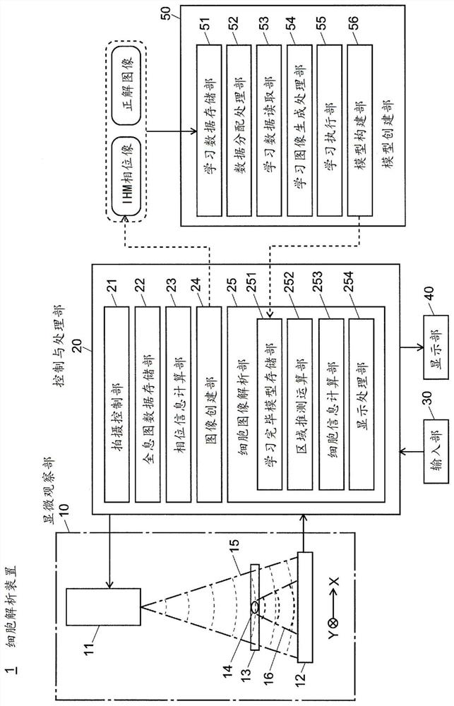
[0064] figure 1 It is a schematic configuration diagram of a cell analysis apparatus using a cell image analysis apparatus for implementing the cell image analysis method of the present invention.

[0065] The cell analysis device 1 of this embodiment includes a microscopic observation unit 10, a control and processing unit 20, an input unit 30 and a display unit 40 as a user interface, and a model creation unit 50.

[0066] The microscopic observation unit 10 is an in-line holographic microscopy (IHM), and includes a light source unit 11 including a laser diode, etc., and an image sensor 12, and cells are arranged between the light source unit 11 and the image sensor 12 The culture dish 13 of the colony (or cell monomer) 14.

[0067] The control and processing unit 20 controls the actions of the microsco...


PUM


Abstract

In the present invention, a phase image is formed by computation from a holographic image of a cell, a segmentation is carried out for each pixel for the phase image using a fully convolutional neuralnetwork, and an undifferentiated cell region, an undifferentiated deviated cell region, a foreign body region, etc. are identified. During learning, when a learning image included in a mini-batch hasbeen read in (S13), the image is randomly inverted vertically or horizontally (S13) and then is rotated by a random angle (S14). A portion of a pre-rotation image which has been lost within the frameby said rotation is compensated for by a mirror-image inversion with an edge of a post-rotation image as an axis thereof (S15). Learning of a fully convolutional neural network is carried out using the learning image thus generated (S16). The same processes are repeated for all mini-batches, and the learning is repeated for a prescribed number of iterations while learning data allocated to the mini-batch is shuffled. Learning model precision is thus improved. As rotationally invariant characteristics may be learned, it is possible to identify, with good precision, cell colonies being of various shapes.

Description

Technical field [0001] The present invention relates to a method and an apparatus for analyzing an observation image obtained by observing cells, and a method for creating a learning model for the analysis. More specifically, it relates to the cultivation of pluripotent stem cells (ES cells or iPS cells). In the process of ), the cell image analysis method, the cell image analysis device, and the learning model creation method are preferable when judging the state of cells in a non-invasive manner or obtaining cell-related information such as the number of cells. Background technique [0002] In recent years, pluripotent stem cells such as iPS cells and ES cells have been widely used for research in the field of regenerative medicine. In the research and development of regenerative medicine using such pluripotent stem cells, it is necessary to cultivate a large number of undifferentiated cells that maintain a pluripotent state. Therefore, it is necessary to select an appropriate...

Claims


Application Information

Patent Timeline
Patent Type & Authority Applications(China)
IPC IPC(8): G06T7/00
CPCG06T7/11G06T7/0012G06T7/62G06T2207/30024G06T2207/20081G06T2207/10056G06T2207/20076G06V20/695G06V10/454G06V10/82G06T2207/20084
Inventor 高桥涉赤泽礼子
Owner SHIMADZU SEISAKUSHO CO LTD
Why Eureka
Try Eureka