Method for quantitatively detecting expression levels of BST1, STAB1 and TLR4 genes and application
A gene expression level, STAB1 technology, applied in the direction of biochemical equipment and methods, microbial measurement/inspection, etc., can solve the problem of poor sensitivity
- Summary
- Abstract
- Description
- Claims
- Application Information
AI Technical Summary
Problems solved by technology
Method used
Image
Examples
Embodiment 1
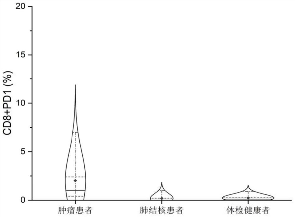
[0027] Example 1 The present invention found that compared with healthy normal people and tuberculosis patients, the peripheral blood CD8+PD-1+T cells of tumor patients are highly expressed.
[0028] Reagents and Materials
[0029] Ficoll-Paque lymphocyte separation medium (GE), red blood cell lysate (BD PMG, catalog number 555899), flow cytometry antibody CD3 APC-H7 (BD PMG, catalog number 560176), CD4 FITC (BD PMG, catalog number 556615), CD8 PerCP (BDIS, Cat. No. 652829), PD1 PE-Cy TM 7 (BD PMG, Cat. No. 561272), where IgG1κPE-Cy TM 7 (BD PMG, Cat. No. 557872) as Isotype Control.
[0030] experimental method:
[0031] Collection and preparation of materials: 248 cases of tumor patients, 620 cases of healthy persons and 272 cases of pulmonary tuberculosis were collected from peripheral blood samples, and 5ml of venous blood was collected and placed in a blood routine tube after anticoagulation with heparin, and then separated with lymphocyte separation fluid. The narrow ...
Embodiment 2
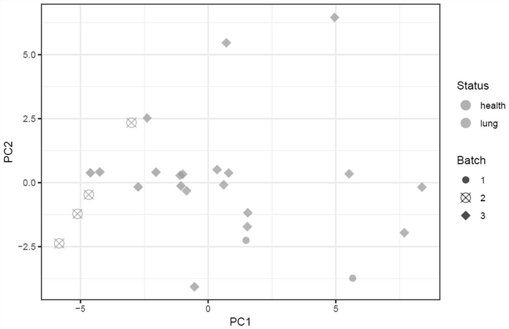
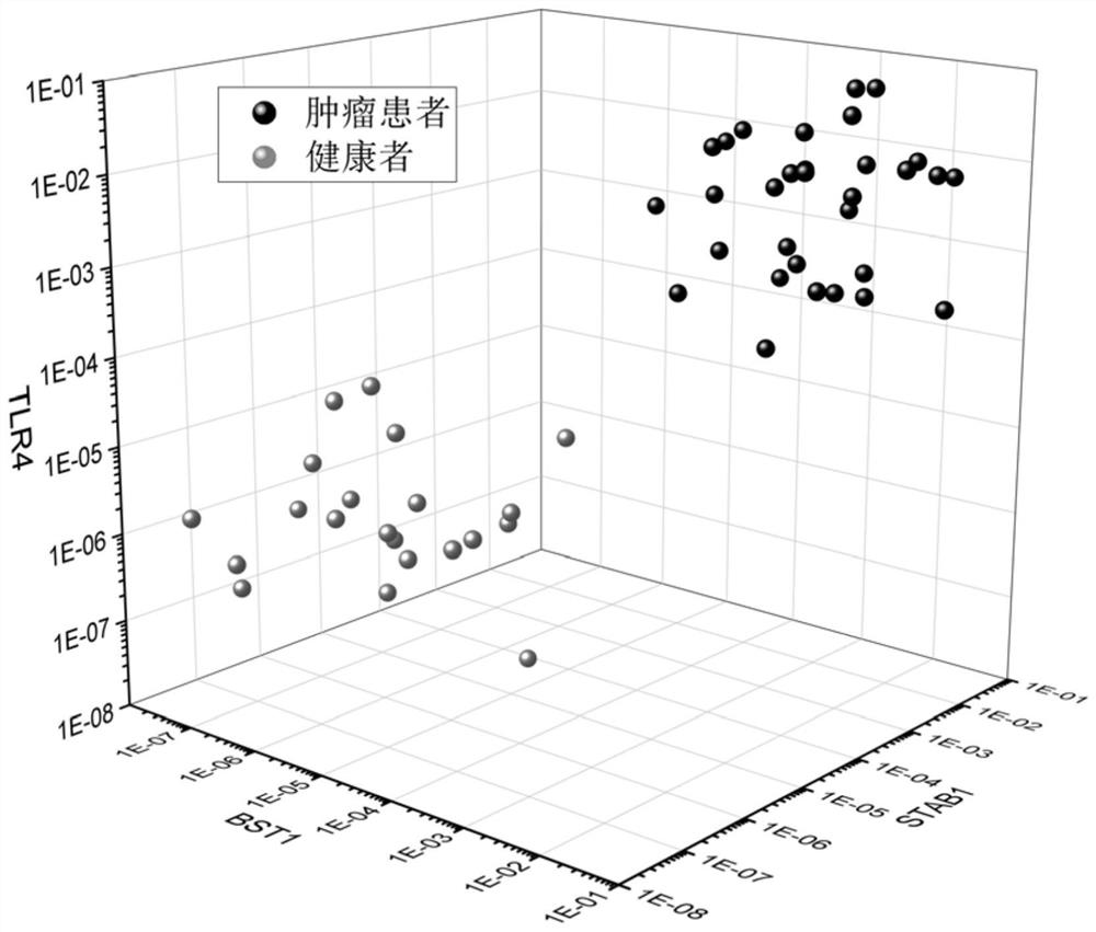
[0035] Example 2 The present invention found that BST1, STAB1 and TLR4 genes were highly expressed in peripheral blood CD8+PD-1+ cells of patients with early lung cancer.
[0036] Reagents and Materials
[0037] See Example 1.
[0038] experimental method:
[0039] Collection and preparation of materials: We selected peripheral blood samples from 17 patients with early lung cancer and 9 normal persons who underwent physical examination in Example 1. The follow-up treatment was the same as in Example 1. 5ml of venous blood was drawn and placed in a blood routine tube after anticoagulation with heparin. , use lymphocyte separation fluid to separate the narrow band of the white cloud layer located at the interface between the upper and middle layers, that is, mononuclear cells (including lymphocytes and monocytes). After being treated with erythrocyte lysate, the pellet was resuspended with PBS, and 20ul was taken for cell counting.
[0040] Flow cytometry antibody staining an...
Embodiment 3
[0054] Example 3 RT-qPCR verification of expression levels of 14 differential genes in lung cancer CD3+CD8+PD1+ cells
[0055] Reagents and Materials
[0056] The peripheral blood of 31 cases of lung cancer, 4 cases of pulmonary granulomatous inflammation and 17 cases of pulmonary tuberculosis were collected. The collection standard is shown in Example 1.
[0057] experimental method
[0058] See Example 1 for the processing of peripheral blood, flow cytometry antibody staining and on-machine detection, and obtain CD3+CD8+PD1+ cells after flow cytometry sorting.
[0059] RNA extraction
[0060] Extract total RNA according to the standard operation of Qiagen RNeasy Micro Kit. After sorting, add Buffer RLT containing 350 μl β-ME and 350 μL 70% ethanol to the cells, shake vigorously and mix well, pass through MinElute filter column, centrifuge at ≥10,000 rpm for 15 seconds, discard waste liquid. Add 350μL Buffer RW1, centrifuge at ≥10000rpm for 15s, and discard the waste. Ad...
PUM
 Abstract
Description
Claims
Application Information
 - R&D
- Intellectual Property
- Life Sciences
- Materials
- Tech Scout
- Unparalleled Data Quality
- Higher Quality Content
- 60% Fewer Hallucinations
Browse by: Latest US Patents, China's latest patents, Technical Efficacy Thesaurus, Application Domain, Technology Topic, Popular Technical Reports.
© 2025 PatSnap. All rights reserved.Legal|Privacy policy|Modern Slavery Act Transparency Statement|Sitemap|About US| Contact US: help@patsnap.com


