Indocyanine green fluorescence combined multimodal image-guided liver portal vein watershed developing method

A technology of indocyanine green and developing method, applied in the field of medical imaging, can solve the problems of inaccurate search, low success rate of ICG fluorescence staining of portal vein drainage, and difficulty in understanding the anatomical structure of ultrasonic images, so as to reduce surgical complications, Improve the accuracy and safety of surgery and improve the effect of treatment efficacy

Pending Publication Date: 2020-11-06
ZHONGSHAN HOSPITAL FUDAN UNIV
View PDF0 Cites 3 Cited by
  • Summary
  • Abstract
  • Description
  • Claims
  • Application Information

AI Technical Summary

Problems solved by technology

Therefore, accurately finding and puncturing the target portal vein branch is the key to the success of this type of operation; however, due to the extremely high variability of the portal vein branch in the liver, the number of branches, the location of the bifurcation, the direction of travel, and the relative position to the tumor are different for each patient. , while the current intraoperative ultrasound is a two-dimensional image, it is difficult for inexperienced doctors to understand the anatomical structure corresponding to the ultrasound image, resulting in the inability to accurately find and puncture the target portal vein branch, which leads to the current intraoperative ultrasound-guided ICG fluorescence staining Poor success rate in portal drainage

Method used


Image

  • Indocyanine green fluorescence combined multimodal image-guided liver portal vein watershed developing method

Examples


Embodiment Construction

[0020] In order to make the present invention more obvious and understandable, the preferred embodiments are described in detail as follows in conjunction with the accompanying drawings:

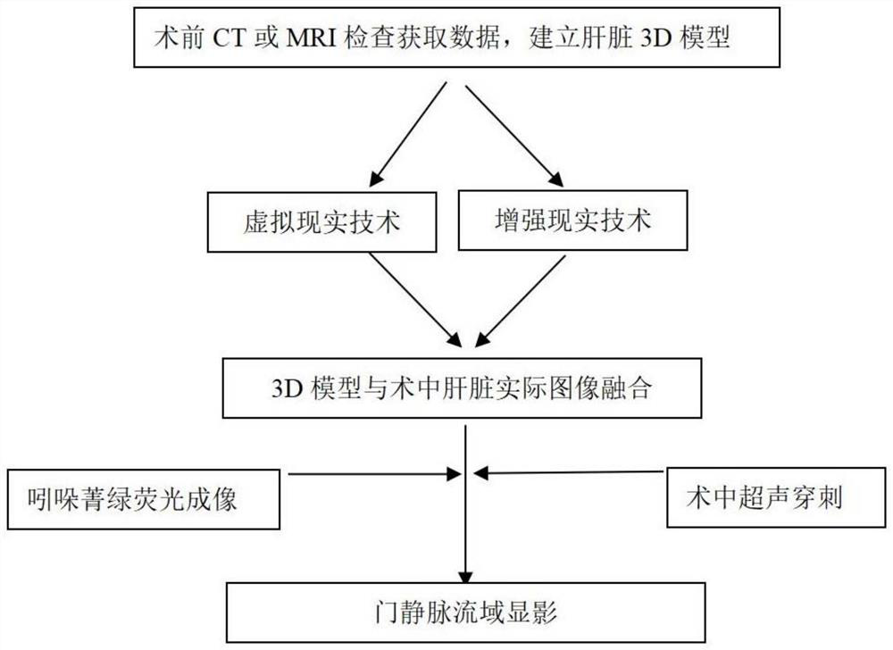
[0021] Such as figure 1 As shown, the present invention provides a method of indocyanine green fluorescence combined with multimodal image-guided liver portal vein development; comprising the following steps:

[0022] Step 1: The patient undergoes preoperative thin-section CT or MRI examination, and uses preoperative 3D visualization software to establish a 3D model of the anatomical structure of the liver and the tumor according to the examination data;

[0023] Step 2: Use virtual reality technology and augmented reality technology to register and fuse the 3D model of the liver anatomical structure generated by the above inspection data with the actual anatomical position of the liver during the operation;

[0024] Step 3: Use fused images to analyze the relationship between the tumor and...


PUM


Abstract

The invention relates to an indocyanine green fluorescence combined multimodal image-guided liver portal vein watershed developing method, and belongs to the technical field of medical images. According to the method, artificial intelligence technologies such as virtual reality, augmented reality and the like are integrated with traditional intraoperative ultrasound and indocyanine green fluorescent staining technologies, so that a surgeon can visualize three-dimensionally complex liver anatomical structures, accurately locate tumors in an operation, plan an operation scheme, accurately achieve developing of a target portal vein watershed, monitor and correct a liver parenchyma disconnection plane in real time, and perform accurate anatomical liver resection. In a liver operation, the method can display a portal vein branch blood flow supply area for supplying tumors in real time, accurately guide a liver resection range, can not only remove micrometastatic tumors that may exist in theportal vein, but also can ensure blood supply of the remaining liver, improve accuracy and safety of the operation, reduce operative complications, and improve efficacy of tumor operation.

Description

technical field [0001] The invention relates to a liver portal vein development method guided by indocyanine green fluorescence combined with multimodal images, which belongs to the technical field of medical imaging. Background technique [0002] Portal vein invasion is an important biological characteristic of hepatocellular carcinoma. In clinical treatment, the purpose of anatomic hepatectomy to resect the portal vein basin of the tumor-bearing liver segment can remove possible residual micrometastases and improve the oncological efficacy of hepatocellular carcinoma. In addition, if the liver parenchyma is cut along the anatomical gap of the liver lobe or liver segment during the operation, there will be less damage to the pipeline during the operation, the time will be faster, the bleeding will be less, and the postoperative complication rate such as bile leakage will be reduced. [0003] Indocyanine green fluorescence (ICG) is a near-infrared fluorescent dye, which can...

Claims


Application Information

Patent Timeline
Patent Type & Authority Applications(China)
IPC IPC(8): A61B5/00A61B17/34A61B34/10A61B34/20A61B90/00A61M5/00
CPCA61B5/0071A61B5/4887A61B5/489A61B17/3403A61B2017/3413A61M5/007A61M2210/1071A61M2210/12A61B34/10A61B34/20A61B90/37A61B2034/107A61B2034/108A61B2034/2055A61B2034/2065A61B2090/365A61B2090/373A61B2090/378A61M2210/005
Inventor 王晓颖
Owner ZHONGSHAN HOSPITAL FUDAN UNIV
Why Eureka
Try Eureka