A dual-enzyme-mediated cascade signal amplification aptasensor for ampicillin detection

An aptamer sensor and ampicillin technology are applied in the determination/inspection of microorganisms, instruments, measuring devices, etc., which can solve the problems of expensive reagents, precision instruments and technicians, low sensitivity and specificity, and cumbersome analysis process, etc., to achieve High binding affinity, strong scalability, and the effect of overcoming steric hindrance

Active Publication Date: 2022-06-24
HUNAN AGRICULTURAL UNIV
View PDF3 Cites 0 Cited by
  • Summary
  • Abstract
  • Description
  • Claims
  • Application Information

AI Technical Summary

Problems solved by technology

[0003] Currently, conventional detection strategies for AMP, such as high-performance liquid chromatography (HPLC), gas chromatography (GC), liquid chromatography-tandem mass spectrometry (HPLC-MS), enzyme-linked immunosorbent assay (ELISA), colorimetry, etc., exist. The disadvantages are low sensitivity and specificity, cumbersome analysis process, expensive reagents, and dependence on precision instruments and technicians.

Method used


Image

  • A dual-enzyme-mediated cascade signal amplification aptasensor for ampicillin detection
  • A dual-enzyme-mediated cascade signal amplification aptasensor for ampicillin detection
  • A dual-enzyme-mediated cascade signal amplification aptasensor for ampicillin detection

Examples


Embodiment 1

[0046] Example 1 Preparation of Probes

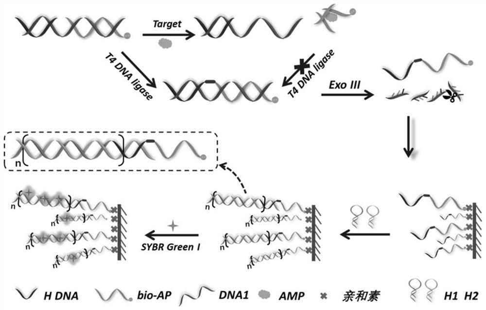
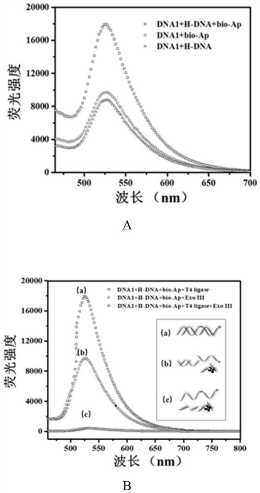
[0047] Take 50 μL of 10 μM DNA1, bio-Apt, and H-DNA priming strands, respectively, and place them in a metal thermostatic oscillator, and activate at 70 °C for 10 min. After mixing evenly, incubate at 4 °C for 2 h to complete the preparation of probes, and store at 4 °C for future use. Then, 10 μL of SYBR Green I with a final concentration of 5× was added to it, incubated at 4 °C for 30 min, and the fluorescence intensity of the sample at 520 nm under excitation light of 490 nm was detected. In order to verify the hybridization between DNA1, bio-Apt and H-DNA. The following samples were selected: a) DNA1 and bio-Apt in equal volume and concentration (10 μL, 10 μM), and 10 μL water; b) DNA1 and H-DNA in equal volume and concentration (10 μL, 10 μM), and 10 μL water. The mixture was incubated at 4 °C for 2 h. Afterwards, 10 μL of SYBR Green I with a final concentration of 5× was added to the two systems respectively, incubated at 4 °C...

Embodiment 2T4

[0049] Example 2 Activity verification of T4 ligase and Exo III

[0050] Test process: a) Take 10 μL of 100 U / mL T4 ligase and 100 μL of detection probe, add 90 μL of PBS buffer solution to mix, incubate at 37 °C for 30 min, and then inactivate at 70 °C for 20 min. After cooling, 10 μL of SYBR Green I with a final concentration of 5× was added and incubated at 4 °C for 30 min. b) Take 10 μL of 100 U / mL Exo III and 100 μL of detection probe, add 90 μL of PBS buffer solution to mix, incubate at 37 °C for 30 min, and then inactivate at 70 °C for 20 min. After cooling, 10 μL of SYBR Green I with a final concentration of 5× was added and incubated at 4 °C for 30 min. c) Take 10 μL of 100U / mL T4 ligase and 100 μL of detection probe, and incubate at 16 °C for 30 min. Add 10 μL 100 U / mL ExoIII and mix with 80 μL PBS buffer solution, incubate at 37 °C for 30 min, and then inactivate at 70 °C for 20 min. After cooling, 10 μL of SYBR Green I with a final concentration of 5× was added ...

Embodiment 3

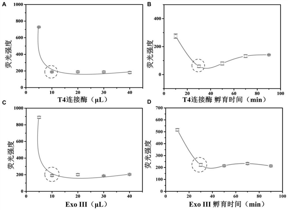
[0052] Example 3 Optimization of experimental conditions for double-enzyme-mediated DNA degradation reaction

[0053] Take 5, 10, 20, 30 and 40 μL of 100 U / mL T4 ligase and 60 μL of detection probe, respectively, and incubate at 16 °C for 30 min. Add 10 μL of 100 U / mL Exo III and mix with 80 μL of PBS buffer, incubate at 37 °C for 30 min, and then inactivate at 70 °C for 20 min. After cooling, 10 μL of SYBR Green I with a final concentration of 5× was added and incubated at 4 °C for 30 min. Detect the fluorescence intensity value of the solution at 520 nm.

[0054] see image 3 A, When the volume of T4 ligase is 10 μL, the fluorescence intensity of the solution reaches a steady state, which is the optimal reaction volume.

[0055] Take 10 μL of 100 U / mL T4 ligase and 60 μL of detection probe, respectively, and incubate at 16 °C for 10, 30, 50, 70 and 90 min. Add 10 μL of 100 U / mL Exo III and mix with 80 μL of PBS buffer, incubate at 37 °C for 30 min, and then inactivate at...


PUM


Abstract

An ampicillin aptamer sensor based on dual-enzyme-mediated cascade signal amplification is prepared by the following steps: using double-stranded DNA formed by mutual hybridization of bio-AP, DNA1 and H-DNA as a detection probe, ligated with T4 DNA With the assistance of enzyme and exonuclease III, the complete APH chain is obtained; through the specific binding of biotin-avidin, the APH chain is captured in the microwell plate as the HCR priming chain, resulting in hairpin DNA H1 and H2 is cross-opened to produce double-stranded DNA polymers, and the fluorescent dye SYBR Green I indicator is added to produce obvious fluorescence when it is embedded in double-stranded DNA. Based on the specific binding of the nucleic acid aptamer to the target, the detection of ampicillin is realized through the change of the fluorescence intensity. The method has high sensitivity, good specificity and high accuracy, and can be applied to the detection of ampicillin in contaminated food such as milk.

Description

technical field [0001] The invention belongs to the technical field of biosensing and nucleic acid detection, and relates to the detection of ampicillin (AMP) in food, in particular to an aptamer sensor for ampicillin detection with double-enzyme-mediated cascade signal amplification. Background technique [0002] As one of the broad-spectrum β-lactam antibiotics, ampicillin, AMP has been widely used in the field of medicine and bacterial infection treatment, and has the advantages of low toxicity, low cost and good clinical effect. In recent years, although AMPs as growth regulators or anti-infectives have promoted the rapid development of animal husbandry and feed processing industries, the illegal and excessive use of AMPs in animals and livestock has directly led to the use of antifungal drugs in the production of human and animal-derived foods. Contamination and residues cause increasingly prominent health and safety issues, such as the emergence of hypersensitivity rea...

Claims


Application Information

Patent Timeline
Patent Type & Authority Patents(China)
IPC IPC(8): C12Q1/682G01N33/53G01N21/64
CPCC12Q1/682G01N33/53G01N21/6428G01N2021/6439
Inventor 石星波赵倩张光胤
Owner HUNAN AGRICULTURAL UNIV
Why Eureka
Try Eureka