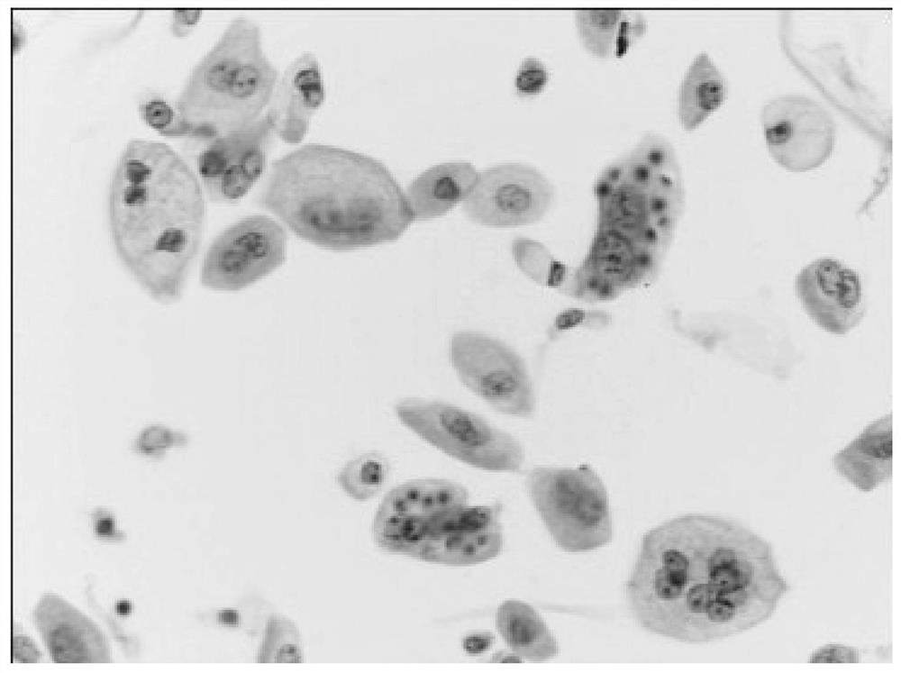
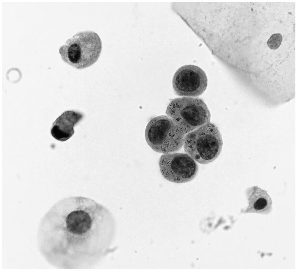
Multi-staining flaking method of cytopathology sample
A pathology and cell technology, applied in the field of cell pathology, can solve the problems of loss, false positive, conventional dye shedding, etc., and achieve the effect of improving accuracy and uniform distribution
- Summary
- Abstract
- Description
- Claims
- Application Information
AI Technical Summary
Problems solved by technology
Method used
Image

Examples
Embodiment 1
[0059] Novel and Improved Immunochemical Protein Marker Staining Method for Cells and Diff Fast Cell Multiplex Staining
[0060] (All centrifugation is performed at 1500 r / min for 5 min, the supernatant is removed, and then PBS is used to prepare the suspension)
[0061] 1. 25ml of bladder cancer patient urine containing 4% paraformaldehyde cell fixative, centrifuged to pellet the cells, discarded the supernatant, then diluted and suspended in 1ml of PBS (pH7.4) buffer, placed in 10ml in plastic test tubes.
[0062] 2. Add 20 microliters of Triton-100 (to increase the permeability of the cell membrane, which is beneficial to the staining of the cytoplasm and nucleus).
[0063] 3. Heat the cell suspension at 92°C for 5 minutes for antigen retrieval.
[0064] 4. Add 100 microliters of Dual Endogenous Enzyme Block (Dako, Carpinteria, Ca) to remove endogenous peroxidase (peroxidase) and alkaline phosphatase (alkline phosphatase) activity in cells, and incubate for 5 minutes.
...
Embodiment 2
[0080] Based on the new and improved staining method of cell nucleic acid chromogenic in situ hybridization and multiple staining of Diff fast cells:
[0081] (All centrifugation is performed at 1500 r / min for 5 min, the supernatant is removed, and then PBS is used to prepare the suspension)
[0082] 1. 25ml of bladder cancer patient urine containing 4% paraformaldehyde cell fixative, centrifuged to pellet the cells, discarded the supernatant, then diluted and suspended in 1ml of PBS (pH7.4) buffer, placed in 10ml in plastic test tubes.
[0083] 2. Add 20 microliters of Triton-100 (to increase the permeability of the cell membrane, which is beneficial to the staining of the cytoplasm and nucleus).
[0084] 3. Purchase RNAscope® 2.5 HD Reagent Kit (ACD Bio, California, USA) immunohistochemical nucleic acid in situ hybridization kit, and perform nucleic acid marker staining according to the experimental procedures recommended by the manufacturer. Add the RNA nucleic acid probe...
PUM
 Abstract
Description
Claims
Application Information