Medical application of cathepsin inhibitor

A technology of cathepsin and inhibitors, applied in the field of medicine, can solve the problems of not being able to meet the growing demand for medication of fibrosis patients

Active Publication Date: 2021-03-05
SHENYANG PHARMA UNIVERSITY
View PDF2 Cites 0 Cited by
  • Summary
  • Abstract
  • Description
  • Claims
  • Application Information

AI Technical Summary

Problems solved by technology

In particular, there are currently only two drugs for the treatment of pulmonary fibrosis

Method used


Image

  • Medical application of cathepsin inhibitor
  • Medical application of cathepsin inhibitor
  • Medical application of cathepsin inhibitor

Examples


Embodiment 1

[0033] Example 1: Cathepsin Inhibitory Activity Test

[0034] 1. Experimental materials: cathepsin L and S at a temperature of 37°C, 5% CO 2 cultured in a humidified incubator. After each compound was dissolved in dimethyl sulfoxide (DMSO) under sterile conditions, it was diluted to the required concentration with RPMI-1640 culture solution, and the final concentration of DMSO was less than 0.5%. RPMI 1640 was purchased from Gibco (Grand Island, USA), fetal bovine serum (FBS) was purchased from Biological Industries, penicillin-streptomycin was purchased from HyClone Company, trypsin (Trypsin, 1:250) was purchased from Biosharp Company, dimethyl Sulfoxide (DMSO) was purchased from Sigma Chemical Company, and monodansylpentamethylenediamide (MDC) was purchased from Nanjing KGI Biotechnology Co., Ltd.

[0035] 2. Instruments: carbon dioxide incubator (SANYO, Japan, model: MCO-5AC), inverted microscope (OLYMPUS, Japan, model: CKX41), enzyme-linked immunoassay analyzer (Tecan, A...

Embodiment 2

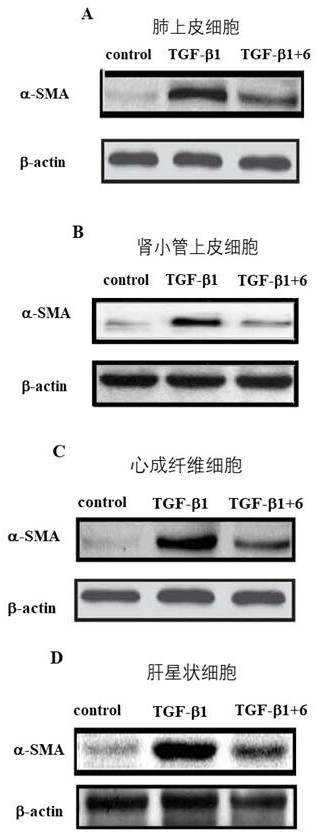
[0056] Example 2: Study on the anti-pulmonary fibrosis, liver fibrosis, kidney fibrosis and cardiac fibrosis of compound 6 based on the fibrosis marker protein α-SMA in vitro.

[0057] Take compound 6 with the best enzyme activity as an example:

[0058] Lung epithelial cells A549 cells, hepatic stellate cells LX-2, renal tubular epithelial cells CD3 and primary cardiac fibroblasts were pre-treated with 300 μM compound 6 or vehicle, and then stimulated by 10 ng / mL TGF-β1 to Myofibroblasts were transformed to construct lung, liver, kidney and heart fibrosis models in vitro, and the fibrosis marker protein α-SMA was used to characterize the degree of fibrosis. like figure 1 As shown, after 10ng / ml TGF-β1 stimulated human lung epithelial cells A549, hepatic stellate cells LX-2, renal tubular epithelial cells CD3 and primary mouse cardiac fibroblasts for 48h, the fibrosis marker protein α-SMA Significantly up-regulated (P<0.01=demonstrates successful construction of classic lung...

Embodiment 3

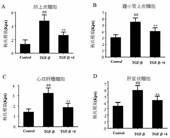
[0059] Example 3: Cell stiffness as a marker of fibrosis The effect of compound 6 on anti-pulmonary fibrosis, liver fibrosis, kidney fibrosis and cardiac fibrosis was investigated in vitro.

[0060] Previous studies have shown that cell stiffness can be used as a biomarker to characterize the degree of fibrosis. Lung epithelial cells A549 cells, hepatic stellate cells LX-2, renal tubular epithelial cells CD3 and primary cardiac fibroblasts were pre-treated with 300 μM compound 6 or vehicle, and then stimulated by 10 ng / mL TGF-β1 to Transformation of myofibroblasts to construct fibrosis models of lung, liver, kidney and heart in vitro. Atomic force microscopy was used to detect the changes in the stiffness of the above four cells after being stimulated by TGF-β1, and the cell stiffness (Young's modulus) was used as a marker to characterize degree of fibrosis. the result shows( figure 2 ), in the above models of pulmonary fibrosis, liver fibrosis, renal fibrosis and cardiac f...


PUM


Abstract

The invention belongs to the technical field of medicine, relates to medical application of cathepsin inhibitors, and specifically relates to application of some cathepsin inhibitors in preparation ofmedicines for preventing or treating fibrotic diseases. The cathepsin inhibitor has a structure shown as follows, wherein R1 and R2 are described in the claims and the specification. The fibrotic diseases comprise pulmonary fibrosis, hepatic fibrosis, renal fibrosis, cardiac fibrosis, endometrial fibrosis, ocular fibrosis, pancreatic fibrosis, spleen fibrosis, myelofibrosis diseases or diseases induced by fibrosis. According to the invention, the cathepsin inhibitor is used alone or used in combination with other drugs.

Description

Technical field: [0001] The invention belongs to the technical field of medicine and relates to the medical application of cathepsin inhibitors, in particular to the application of some cathepsin inhibitors in the preparation of drugs for preventing or treating fibrosis diseases. Background technique: [0002] Fibrosis refers to the pathological process in which inflammation leads to necrosis of organ parenchymal cells and abnormal increase and excessive deposition of extracellular matrix in tissues. Essentially, fibrosis is a repair response to tissue damage to protect the relative integrity of tissues and organs. Although the proliferating fibrous connective tissue repairs the defect, it does not have the structure and function of the original organ parenchymal cells. If this repair response is excessive, strong, and out of control, it will cause organ fibrosis and lead to a decline in organ function. [0003] Fibrosis can occur in many organs, including vital organs suc...

Claims


Application Information

Patent Timeline
IPC IPC(8): A61K31/216A61K31/4164A61K31/4196A61K31/4409A61P11/00A61P1/16A61P13/12A61P15/00A61P27/02A61P1/18A61P19/00A61P37/00A61P9/04A61P37/06A61P3/06A61P3/10A61P19/06A61P9/10A61P9/12A61P31/12A61P3/00A61P43/00
CPCA61K31/216A61K31/4164A61K31/4196A61K31/4409A61P1/16A61P1/18A61P3/00A61P3/06A61P3/10A61P9/04A61P9/10A61P9/12A61P11/00A61P13/12A61P15/00A61P19/00A61P19/06A61P27/02A61P31/12A61P37/00A61P37/06A61P43/00
Inventor 袁雷马恩龙
Owner SHENYANG PHARMA UNIVERSITY
Why Eureka
Try Eureka