

Large-view-field high-flux high-resolution pathological section analyzer
A pathological slice and high-throughput technology, applied in the field of medical imaging, can solve problems such as system complexity, time-consuming, and time-consuming, and achieve the effects of simplifying the analysis process, reducing cost consumption, and avoiding errors
- Summary
- Abstract
- Description
- Claims
- Application Information
AI Technical Summary
Problems solved by technology
Method used
Image

Examples
Embodiment 1
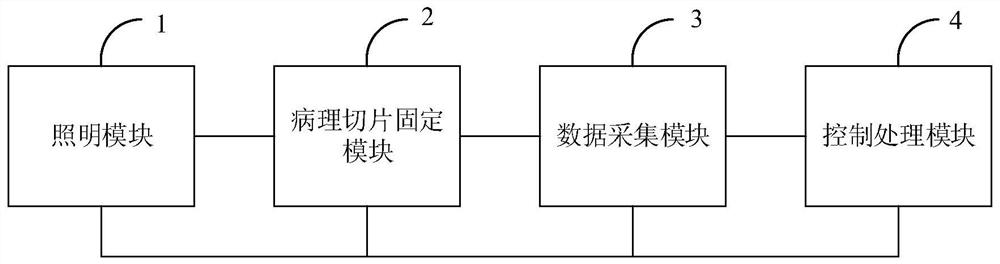
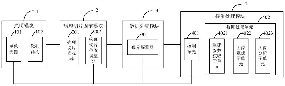
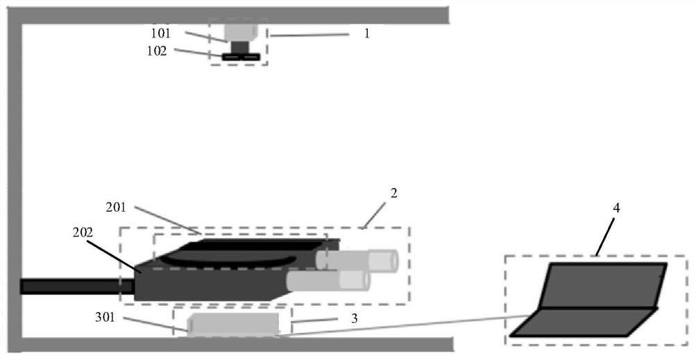
[0051] See figure 1 , figure 1 It is a structural block diagram of a large-field-of-view high-throughput high-resolution pathological slice analyzer provided by an embodiment of the present invention. As shown in the figure, the large-field-of-view high-throughput high-resolution pathological slice analyzer of the embodiment of the present invention includes:
[0052] A lighting module 1, configured to generate monochromatic light;
[0053] Pathological slice fixing module 2, configured to fix and adjust the position of the pathological slice, so that the pathological slice is located at the position of the imaging field of view;
[0054] The data collection module 3 is used to collect the interference image information formed by the scattered light carrying the wavefront information after the monochromatic light passes through the pathological slice, and the transmitted light not carrying the pathological slice information;
[0055] The control processing module 4 is confi...
PUM
 Abstract
Description
Claims
Application Information