

Breast cancer bone metastasis laboratory mouse disease model building method
A disease model and construction method technology, applied in medical science, veterinary instruments, veterinary surgery, etc., can solve the problems of high operation difficulty and low success rate of modeling, so as to improve the success rate of modeling and tumor survival rate effect
- Summary
- Abstract
- Description
- Claims
- Application Information
AI Technical Summary
Problems solved by technology
Method used
Image

Examples
Embodiment 1
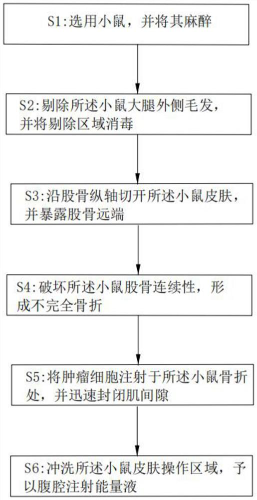
[0039] The method for constructing the experimental mouse disease model of breast cancer bone metastasis in the present embodiment comprises the following steps:
[0040] S1: choose 20g female mice, give 1% ketamine intraperitoneal injection, the anesthesia dose is 4ml / kg;
[0041] S2: After the anesthesia is completed, move to the sterile operating table, fix it in the lateral position, remove about 1cm*1cm of hair on the outer thigh of the mouse along the longitudinal axis of the femur, and the removed area coincides with the femur, and disinfect it with 1% iodine for 3 times. and wipe clean with sterile gauze;
[0042] S3: Cut the skin 0.8 cm along the longitudinal axis of the femur, bluntly separate the subcutaneous fascia, and bluntly separate the muscle space between the lateral rectus femoris and semitendinosus with a small hemostat to expose the distal end of the femur (pay attention to protection during separation) medial femoral artery located in the femur);
[004...
Embodiment 2
[0048] The method for constructing the experimental mouse disease model of breast cancer bone metastasis in the present embodiment comprises the following steps:
[0049] S1: choose 20g female mice, give 1% ketamine intraperitoneal injection, the anesthesia dose is 4ml / kg;
[0050] S2: After the anesthesia is completed, move to the sterile operating table, fix it in the lateral position, remove about 1cm*1cm of hair on the outer thigh of the mouse along the longitudinal axis of the femur, and the removed area coincides with the femur, and disinfect it with 1% iodine for 3 times. and wipe clean with sterile gauze;
[0051] S3: Cut the skin 0.8 cm along the longitudinal axis of the femur, bluntly separate the subcutaneous fascia, and bluntly separate the muscle space between the lateral rectus femoris and semitendinosus with a small hemostat to expose the distal end of the femur (pay attention to protection during separation) medial femoral artery located in the femur);
[005...
Embodiment 3
[0064] The method for constructing the experimental mouse disease model of breast cancer bone metastasis in the present embodiment comprises the following steps:
[0065] S1: choose 25g female mice, give 1% ketamine intraperitoneal injection, the anesthesia dose is 4ml / kg;
[0066] S2: After the anesthesia is complete, move to the sterile operating table, fix in the lateral position, and remove about 1.5cm*1.5cm of hair on the outer thigh of the mouse along the longitudinal axis of the femur. and wipe clean with sterile gauze;
[0067] S3: Cut the skin 0.8 cm along the longitudinal axis of the femur, bluntly separate the subcutaneous fascia, and bluntly separate the muscle space between the lateral rectus femoris and semitendinosus with a small hemostat to expose the distal end of the femur (pay attention to protection during separation) medial femoral artery located in the femur);
[0068] S4: Use tooth forceps to clamp the proximal 1 / 3 of the femur, ophthalmic scissors to ...
PUM
| Property | Measurement | Unit |
|---|---|---|
| Length | aaaaa | aaaaa |
Abstract
Description
Claims
Application Information