
High-definition PET image reconstruction method

An image reconstruction, high-definition technology, applied in the field of medical image image processing, can solve the problems of ignoring inter-crystal scattering, reducing image resolution and contrast, and low accuracy, reducing storage space and computational complexity, and improving image signal-to-noise. Ratio and contrast, the effect of improving accuracy

Pending Publication Date: 2021-07-13
XUZHOU MEDICAL UNIV
View PDF0 Cites 1 Cited by
  • Summary
  • Abstract
  • Description
  • Claims
  • Application Information

AI Technical Summary

Problems solved by technology

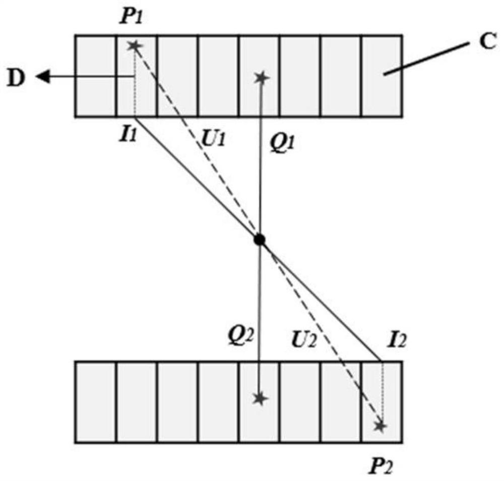
[0005] PSF modeling mainly includes three methods: analytical model, Monte Carlo simulation (monte carlo, MC), and experimental method. The analytical model is mainly an analytical model of the penetration between crystals determined by the incident angle, so that a single detector can be blurred. Extended to meet the radial ambiguity of the detector pair, however, this method ignores the contribution of inter-crystal scattering and only considers the inter-crystal penetration, which is less accurate than the other two methods; the MC simulation PSF response function only estimates any given The ambiguity between the radial direction and the azimuth angle in a given direct or oblique two-dimensional sinogram; using experimental methods to obtain the spatially varying PSF values ​​​​of voxels at each fixed point position within the PET system's field of view is a huge task, which is not only time-consuming and expensive. Serious waste of computer storage resources, and the analysis and processing of later experimental data is also extremely challenging
[0006] In order to solve the problem that the resolution and contrast of the reconstructed image are reduced due to the depth effect between crystals, which affects the high-precision identification of tiny lesions, a more accurate modeling of the PET system is required, that is, high-definition PET image reconstruction

Method used


Image

  • High-definition PET image reconstruction method
  • High-definition PET image reconstruction method
  • High-definition PET image reconstruction method

Examples


Embodiment Construction

[0053] The present invention will be further described below.

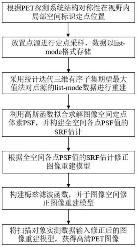
[0054] Such as figure 1 Shown, a kind of high-definition PET image reconstruction method comprises the following steps:

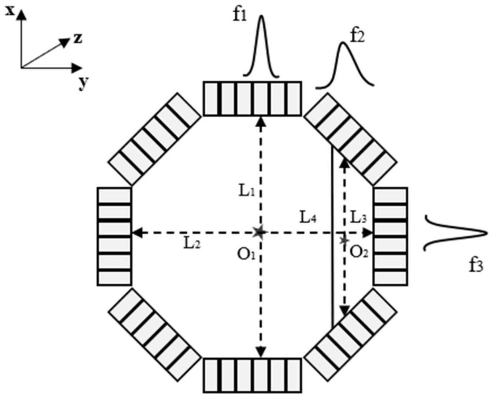
[0055] ① According to the structural symmetry of the PET detection system, the fixed-point position is marked in the local space within the field of vision;

[0056] ② Place a point source at a fixed point for sampling at a fixed point, and the data is stored in list-mode format;

[0057] ③Reconstruct the list-mode data of the point source by using the statistical iterative three-dimensional ordered subset expected maximum method;

[0058] ④Use Gaussian function fitting to solve the PSF value of each fixed point voxel in the image space, and construct the SRF estimation of the PSF value of each fixed point in the whole space;

[0059] ⑤ Correct the image reconstruction model according to the SRF estimation of the PSF value of each fixed point in the whole space;

[0060] ⑥Construct the Met...


PUM


Abstract

The invention discloses a high-definition PET image reconstruction method. The method comprises the following steps: marking a fixed point position in a local space in a visual field according to structural symmetry of a PET detection system; placing a point source for fixed-point sampling; reconstructing the list-mode data of the point source by adopting a three-dimensional ordered subset expectation maximum value method; fitting and solving an image space fixed-point voxel point diffusion function value by using a Gaussian function, and constructing system response function estimation of each point in the whole space; estimating a corrected image reconstruction model according to a system response function; constructing a Metz filtering function, and correcting an image reconstruction model in an image space; and inputting actual measurement data of a scanned object into the corrected image reconstruction model to obtain a high-definition PET image. Accurate system modeling is carried out on the list-mode data based on the point spread function, the experiment duration and the data storage space required by system response function modeling are reduced, the calculation complexity is reduced, filtering processing is carried out by using the Mitz filtering function, and the signal-to-noise ratio and the contrast ratio of the image are improved.

Description

technical field [0001] The invention relates to an image reconstruction method, in particular to a high-definition PET image reconstruction method, and belongs to the technical field of medical image processing. Background technique [0002] Positron Emission Tomography (hereinafter referred to as PET) is an advanced nuclear medicine imaging device used in preclinical research and clinical practice to diagnose and guide treatment. It has been widely used in tumors, cardiovascular diseases and nervous system diseases. etc. [0003] From the physical process of positron annihilation, to the detector detection process and electronic readout system, to the image reconstruction algorithm, these processes involve the influence of different factors (including the positron emission stage: positron range, gamma photon non-collinearity , tissue attenuation characteristics of photons, Compton scattering of photons, etc.; photon detection stage: system dead time, detection efficiency a...

Claims


Application Information

Patent Timeline
Patent Type & Authority Applications(China)
IPC IPC(8): G06T11/00A61B6/00A61B6/03
CPCG06T11/006A61B6/037A61B6/52G06T2211/424G06T2211/416
Inventor 刘琼李超凡陈志成赵杰
Owner XUZHOU MEDICAL UNIV
Learn More